Atomistry » Iron » PDB 1yqp-1zj9 » 1zj9
Atomistry »
  Iron »
    PDB 1yqp-1zj9 »
      1zj9 »

Iron in PDB 1zj9: Structure of Mycobacterium Tuberculosis Nira Protein

Enzymatic activity of Structure of Mycobacterium Tuberculosis Nira Protein

All present enzymatic activity of Structure of Mycobacterium Tuberculosis Nira Protein:
1.7.7.1;

Protein crystallography data

The structure of Structure of Mycobacterium Tuberculosis Nira Protein, PDB code: 1zj9 was solved by R.Schnell, T.Sandalova, U.Hellman, Y.Lindqvist, G.Schneider, with X-Ray Crystallography technique. A brief refinement statistics is given in the table below:

Resolution Low / High (Å) 55.00 / 2.90
Space group P 1 21 1
Cell size a, b, c (Å), α, β, γ (°) 59.983, 83.307, 108.801, 90.00, 102.22, 90.00
R / Rfree (%) 20.2 / 28

Other elements in 1zj9:

The structure of Structure of Mycobacterium Tuberculosis Nira Protein also contains other interesting chemical elements:

Chlorine (Cl) 2 atoms

Iron Binding Sites:

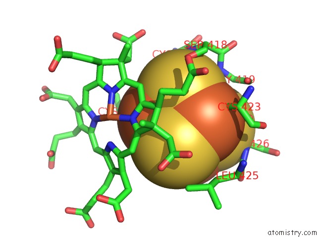
The binding sites of Iron atom in the Structure of Mycobacterium Tuberculosis Nira Protein (pdb code 1zj9). This binding sites where shown within 5.0 Angstroms radius around Iron atom.
In total 10 binding sites of Iron where determined in the Structure of Mycobacterium Tuberculosis Nira Protein, PDB code: 1zj9:
Jump to Iron binding site number: 1; 2; 3; 4; 5; 6; 7; 8; 9; 10;

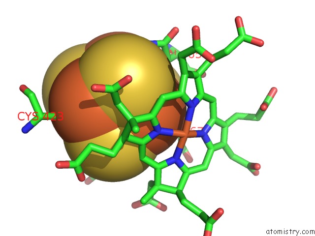
Iron binding site 1 out of 10 in 1zj9

Go back to Iron Binding Sites List in 1zj9
Iron binding site 1 out of 10 in the Structure of Mycobacterium Tuberculosis Nira Protein


Mono view


Stereo pair view

A full contact list of Iron with other atoms in the Fe binding site number 1 of Structure of Mycobacterium Tuberculosis Nira Protein within 5.0Å range:
probe atom residue distance (Å) B Occ
A:Fe556

b:22.0
occ:1.00
FE1 A:SF4556 0.0 22.0 1.0
S4 A:SF4556 2.3 20.1 1.0
S2 A:SF4556 2.3 20.8 1.0
S3 A:SF4556 2.3 24.0 1.0
SG A:CYS463 2.3 27.9 1.0
FE2 A:SF4556 2.6 19.6 1.0
FE3 A:SF4556 2.7 21.5 1.0
FE4 A:SF4556 2.7 20.6 1.0
N A:CYS463 3.4 29.1 1.0
CB A:CYS463 3.5 28.2 1.0
S1 A:SF4556 3.8 23.7 1.0
CA A:CYS463 3.9 28.4 1.0
CB A:ASN465 3.9 26.3 1.0
ND2 A:ASN465 4.0 26.4 1.0
C A:GLY462 4.2 29.8 1.0
O A:CYS463 4.2 27.8 1.0
C A:CYS463 4.2 28.1 1.0
CA A:GLY462 4.4 30.2 1.0
CG A:ASN465 4.5 26.8 1.0
CB A:SER426 4.5 34.5 1.0
N A:GLY462 4.6 30.8 1.0
SG A:CYS423 4.6 30.5 1.0
N A:ASN465 4.6 26.6 1.0
SG A:CYS467 4.7 22.0 1.0
CA A:SER426 4.8 34.4 1.0
CA A:ASN465 4.8 26.3 1.0
SG A:CYS417 4.9 27.1 1.0
O A:LEU425 5.0 33.0 1.0

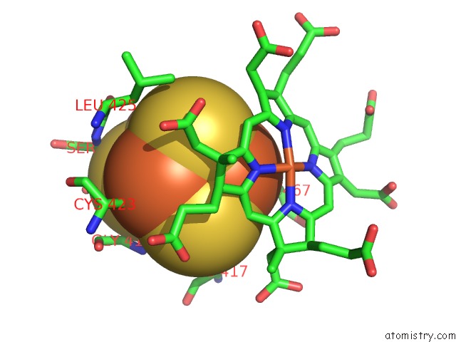
Iron binding site 2 out of 10 in 1zj9

Go back to Iron Binding Sites List in 1zj9
Iron binding site 2 out of 10 in the Structure of Mycobacterium Tuberculosis Nira Protein


Mono view


Stereo pair view

A full contact list of Iron with other atoms in the Fe binding site number 2 of Structure of Mycobacterium Tuberculosis Nira Protein within 5.0Å range:
probe atom residue distance (Å) B Occ
A:Fe556

b:19.6
occ:1.00
FE2 A:SF4556 0.0 19.6 1.0
SG A:CYS467 2.3 22.0 1.0
S4 A:SF4556 2.3 20.1 1.0
S3 A:SF4556 2.3 24.0 1.0
S1 A:SF4556 2.3 23.7 1.0
FE1 A:SF4556 2.6 22.0 1.0
FE3 A:SF4556 2.7 21.5 1.0
FE4 A:SF4556 2.8 20.6 1.0
CB A:CYS467 3.0 23.8 1.0
C4A A:SRM569 3.9 39.1 1.0
S2 A:SF4556 3.9 20.8 1.0
NA A:SRM569 3.9 38.5 1.0
CHB A:SRM569 4.0 37.0 1.0
CA A:CYS467 4.2 23.7 1.0
N A:CYS467 4.2 24.5 1.0
CB A:ASN465 4.3 26.3 1.0
C1A A:SRM569 4.3 39.8 1.0
C3A A:SRM569 4.4 40.4 1.0
CB A:CYS417 4.4 29.1 1.0
FE A:SRM569 4.5 36.7 1.0
C1B A:SRM569 4.6 34.6 1.0
SG A:CYS423 4.6 30.5 1.0
SG A:CYS417 4.7 27.1 1.0
SG A:CYS463 4.7 27.9 1.0
NB A:SRM569 4.8 35.2 1.0
CHA A:SRM569 4.8 39.3 1.0
ND A:SRM569 4.8 37.4 1.0
ND2 A:ASN465 4.8 26.4 1.0
C2A A:SRM569 4.9 40.7 1.0
CDA A:SRM569 4.9 42.6 1.0
C4D A:SRM569 4.9 38.5 1.0

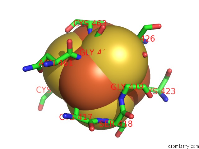
Iron binding site 3 out of 10 in 1zj9

Go back to Iron Binding Sites List in 1zj9
Iron binding site 3 out of 10 in the Structure of Mycobacterium Tuberculosis Nira Protein


Mono view


Stereo pair view

A full contact list of Iron with other atoms in the Fe binding site number 3 of Structure of Mycobacterium Tuberculosis Nira Protein within 5.0Å range:
probe atom residue distance (Å) B Occ
A:Fe556

b:21.5
occ:1.00
FE3 A:SF4556 0.0 21.5 1.0
S2 A:SF4556 2.3 20.8 1.0
S1 A:SF4556 2.3 23.7 1.0
SG A:CYS423 2.3 30.5 1.0
S4 A:SF4556 2.3 20.1 1.0
FE1 A:SF4556 2.7 22.0 1.0
FE2 A:SF4556 2.7 19.6 1.0
FE4 A:SF4556 2.7 20.6 1.0
CB A:CYS423 3.2 30.6 1.0
S3 A:SF4556 3.9 24.0 1.0
CB A:SER426 4.0 34.5 1.0
N A:GLY419 4.3 30.8 1.0
C3A A:SRM569 4.4 40.4 1.0
CDA A:SRM569 4.5 42.6 1.0
SG A:CYS463 4.6 27.9 1.0
CA A:GLY419 4.6 30.9 1.0
CA A:CYS423 4.7 30.9 1.0
SG A:CYS417 4.7 27.1 1.0
N A:SER426 4.7 34.0 1.0
C4A A:SRM569 4.8 39.1 1.0
SG A:CYS467 4.8 22.0 1.0
CA A:SER426 4.8 34.4 1.0
CB A:LEU425 4.9 32.7 1.0
CBA A:SRM569 5.0 41.4 1.0

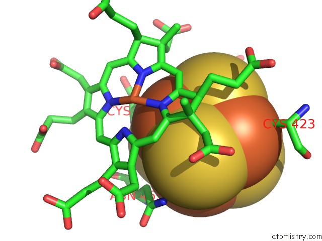
Iron binding site 4 out of 10 in 1zj9

Go back to Iron Binding Sites List in 1zj9
Iron binding site 4 out of 10 in the Structure of Mycobacterium Tuberculosis Nira Protein


Mono view


Stereo pair view

A full contact list of Iron with other atoms in the Fe binding site number 4 of Structure of Mycobacterium Tuberculosis Nira Protein within 5.0Å range:
probe atom residue distance (Å) B Occ
A:Fe556

b:20.6
occ:1.00
FE4 A:SF4556 0.0 20.6 1.0
S1 A:SF4556 2.2 23.7 1.0
SG A:CYS417 2.3 27.1 1.0
S2 A:SF4556 2.3 20.8 1.0
S3 A:SF4556 2.3 24.0 1.0
FE3 A:SF4556 2.7 21.5 1.0
FE1 A:SF4556 2.7 22.0 1.0
FE2 A:SF4556 2.8 19.6 1.0
CB A:CYS417 3.0 29.1 1.0
N A:GLY462 3.3 30.8 1.0
CA A:GLY462 3.6 30.2 1.0
N A:GLY419 3.9 30.8 1.0
S4 A:SF4556 4.0 20.1 1.0
N A:CYS463 4.1 29.1 1.0
CA A:GLY419 4.3 30.9 1.0
N A:SER418 4.3 30.1 1.0
CB A:CYS467 4.3 23.8 1.0
C A:GLY462 4.3 29.8 1.0
CA A:CYS417 4.4 29.4 1.0
C A:ASN461 4.5 31.2 1.0
SG A:CYS467 4.6 22.0 1.0
C A:CYS417 4.7 29.8 1.0
CB A:SER426 4.8 34.5 1.0
CA A:ASN461 4.8 31.4 1.0
SG A:CYS463 4.9 27.9 1.0
SG A:CYS423 4.9 30.5 1.0
C A:SER418 5.0 30.8 1.0

Iron binding site 5 out of 10 in 1zj9

Go back to Iron Binding Sites List in 1zj9
Iron binding site 5 out of 10 in the Structure of Mycobacterium Tuberculosis Nira Protein


Mono view


Stereo pair view

A full contact list of Iron with other atoms in the Fe binding site number 5 of Structure of Mycobacterium Tuberculosis Nira Protein within 5.0Å range:
probe atom residue distance (Å) B Occ
A:Fe569

b:36.7
occ:1.00
FE A:SRM569 0.0 36.7 1.0
NC A:SRM569 1.9 33.6 1.0
NB A:SRM569 2.0 35.2 1.0
NA A:SRM569 2.0 38.5 1.0
ND A:SRM569 2.0 37.4 1.0
CL A:CL570 2.6 34.4 1.0
SG A:CYS467 2.6 22.0 1.0
C1C A:SRM569 2.9 31.2 1.0
C4B A:SRM569 2.9 33.8 1.0
C1D A:SRM569 3.0 37.0 1.0
C4A A:SRM569 3.0 39.1 1.0
C4C A:SRM569 3.0 34.8 1.0
C4D A:SRM569 3.0 38.5 1.0
C1B A:SRM569 3.0 34.6 1.0
C1A A:SRM569 3.0 39.8 1.0
CHC A:SRM569 3.3 31.7 1.0
CHB A:SRM569 3.3 37.0 1.0
CHA A:SRM569 3.4 39.3 1.0
CHD A:SRM569 3.5 36.2 1.0
NZ A:LYS207 3.8 25.4 1.0
CB A:CYS467 4.0 23.8 1.0
C3C A:SRM569 4.1 33.3 1.0
C2C A:SRM569 4.1 32.9 1.0
C2D A:SRM569 4.1 38.3 1.0
C3D A:SRM569 4.2 37.6 1.0
C3B A:SRM569 4.2 32.5 1.0
NH2 A:ARG97 4.3 24.3 1.0
C3A A:SRM569 4.3 40.4 1.0
C2A A:SRM569 4.3 40.7 1.0
C2B A:SRM569 4.3 33.2 1.0
FE2 A:SF4556 4.5 19.6 1.0
CA A:CYS467 4.6 23.7 1.0
CAB A:SRM569 4.6 32.3 1.0
NZ A:LYS209 4.7 13.9 1.0
CMA A:SRM569 4.9 40.0 1.0

Iron binding site 6 out of 10 in 1zj9

Go back to Iron Binding Sites List in 1zj9
Iron binding site 6 out of 10 in the Structure of Mycobacterium Tuberculosis Nira Protein


Mono view


Stereo pair view

A full contact list of Iron with other atoms in the Fe binding site number 6 of Structure of Mycobacterium Tuberculosis Nira Protein within 5.0Å range:
probe atom residue distance (Å) B Occ
B:Fe1556

b:29.5
occ:1.00
FE1 B:SF41556 0.0 29.5 1.0
SG B:CYS463 2.2 31.8 1.0
S4 B:SF41556 2.3 27.8 1.0
S2 B:SF41556 2.3 28.9 1.0
S3 B:SF41556 2.3 28.8 1.0
FE2 B:SF41556 2.6 26.5 1.0
FE3 B:SF41556 2.7 28.0 1.0
FE4 B:SF41556 2.8 24.0 1.0
N B:CYS463 3.4 30.7 1.0
CB B:CYS463 3.4 30.5 1.0
S1 B:SF41556 3.9 26.6 1.0
CA B:CYS463 3.9 30.4 1.0
CB B:ASN465 3.9 27.0 1.0
O B:CYS463 4.2 30.2 1.0
C B:GLY462 4.2 31.1 1.0
C B:CYS463 4.3 30.0 1.0
ND2 B:ASN465 4.3 25.6 1.0
SG B:CYS467 4.4 26.4 1.0
CA B:GLY462 4.4 31.3 1.0
CG B:ASN465 4.5 26.4 1.0
N B:GLY462 4.5 31.5 1.0
SG B:CYS423 4.6 31.8 1.0
N B:ASN465 4.6 27.8 1.0
CA B:ASN465 4.8 27.2 1.0
CB B:LEU425 4.9 33.8 1.0
N B:SER466 4.9 26.8 1.0
SG B:CYS417 4.9 29.0 1.0
CA B:SER426 5.0 35.6 1.0
N B:SER426 5.0 35.2 1.0

Iron binding site 7 out of 10 in 1zj9

Go back to Iron Binding Sites List in 1zj9
Iron binding site 7 out of 10 in the Structure of Mycobacterium Tuberculosis Nira Protein


Mono view


Stereo pair view

A full contact list of Iron with other atoms in the Fe binding site number 7 of Structure of Mycobacterium Tuberculosis Nira Protein within 5.0Å range:
probe atom residue distance (Å) B Occ
B:Fe1556

b:26.5
occ:1.00
FE2 B:SF41556 0.0 26.5 1.0
S3 B:SF41556 2.3 28.8 1.0
SG B:CYS467 2.3 26.4 1.0
S4 B:SF41556 2.3 27.8 1.0
S1 B:SF41556 2.3 26.6 1.0
FE1 B:SF41556 2.6 29.5 1.0
FE4 B:SF41556 2.7 24.0 1.0
FE3 B:SF41556 2.7 28.0 1.0
CB B:CYS467 2.9 25.5 1.0
S2 B:SF41556 3.9 28.9 1.0
NA B:SRM1569 3.9 38.5 1.0
C4A B:SRM1569 3.9 39.0 1.0
CHB B:SRM1569 4.1 36.3 1.0
CA B:CYS467 4.1 25.5 1.0
N B:CYS467 4.2 25.8 1.0
CB B:CYS417 4.3 29.9 1.0
SG B:CYS417 4.4 29.0 1.0
C3A B:SRM1569 4.4 40.1 1.0
C1A B:SRM1569 4.4 39.5 1.0
SG B:CYS423 4.5 31.8 1.0
FE B:SRM1569 4.6 33.5 1.0
C1B B:SRM1569 4.7 34.0 1.0
SG B:CYS463 4.7 31.8 1.0
NB B:SRM1569 4.7 34.9 1.0
CB B:ASN465 4.8 27.0 1.0
ND B:SRM1569 4.9 37.2 1.0
C2A B:SRM1569 5.0 40.6 1.0
CHA B:SRM1569 5.0 38.8 1.0

Iron binding site 8 out of 10 in 1zj9

Go back to Iron Binding Sites List in 1zj9
Iron binding site 8 out of 10 in the Structure of Mycobacterium Tuberculosis Nira Protein


Mono view


Stereo pair view

A full contact list of Iron with other atoms in the Fe binding site number 8 of Structure of Mycobacterium Tuberculosis Nira Protein within 5.0Å range:
probe atom residue distance (Å) B Occ
B:Fe1556

b:28.0
occ:1.00
FE3 B:SF41556 0.0 28.0 1.0
S2 B:SF41556 2.3 28.9 1.0
S1 B:SF41556 2.3 26.6 1.0
S4 B:SF41556 2.3 27.8 1.0
SG B:CYS423 2.3 31.8 1.0
FE1 B:SF41556 2.7 29.5 1.0
FE4 B:SF41556 2.7 24.0 1.0
FE2 B:SF41556 2.7 26.5 1.0
CB B:CYS423 3.2 32.2 1.0
S3 B:SF41556 3.9 28.8 1.0
OG B:SER426 4.0 36.7 1.0
CB B:SER426 4.3 35.8 1.0
N B:SER426 4.4 35.2 1.0
N B:GLY419 4.4 31.4 1.0
SG B:CYS417 4.4 29.0 1.0
CA B:GLY419 4.5 31.8 1.0
SG B:CYS463 4.6 31.8 1.0
CA B:SER426 4.6 35.6 1.0
CA B:CYS423 4.7 32.5 1.0
C3A B:SRM1569 4.7 40.1 1.0
CDA B:SRM1569 4.8 42.5 1.0
O B:GLY419 4.8 32.0 1.0
CB B:LEU425 4.9 33.8 1.0
SG B:CYS467 4.9 26.4 1.0
C B:GLY419 5.0 32.1 1.0
C B:LEU425 5.0 34.8 1.0
C4A B:SRM1569 5.0 39.0 1.0
CA B:GLY462 5.0 31.3 1.0
OG B:SER418 5.0 31.2 1.0

Iron binding site 9 out of 10 in 1zj9

Go back to Iron Binding Sites List in 1zj9
Iron binding site 9 out of 10 in the Structure of Mycobacterium Tuberculosis Nira Protein


Mono view


Stereo pair view

A full contact list of Iron with other atoms in the Fe binding site number 9 of Structure of Mycobacterium Tuberculosis Nira Protein within 5.0Å range:
probe atom residue distance (Å) B Occ
B:Fe1556

b:24.0
occ:1.00
FE4 B:SF41556 0.0 24.0 1.0
SG B:CYS417 2.2 29.0 1.0
S1 B:SF41556 2.3 26.6 1.0
S3 B:SF41556 2.3 28.8 1.0
S2 B:SF41556 2.3 28.9 1.0
FE2 B:SF41556 2.7 26.5 1.0
FE3 B:SF41556 2.7 28.0 1.0
FE1 B:SF41556 2.8 29.5 1.0
N B:GLY462 3.0 31.5 1.0
CB B:CYS417 3.2 29.9 1.0
CA B:GLY462 3.4 31.3 1.0
S4 B:SF41556 3.9 27.8 1.0
N B:CYS463 4.0 30.7 1.0
N B:GLY419 4.2 31.4 1.0
C B:GLY462 4.2 31.1 1.0
C B:ASN461 4.2 31.8 1.0
CA B:GLY419 4.3 31.8 1.0
CA B:CYS417 4.5 30.0 1.0
CA B:ASN461 4.6 32.1 1.0
CB B:CYS467 4.6 25.5 1.0
SG B:CYS463 4.7 31.8 1.0
N B:SER418 4.7 30.7 1.0
SG B:CYS467 4.8 26.4 1.0
C B:CYS417 4.9 30.3 1.0
CB B:SER426 4.9 35.8 1.0
CB B:ASN461 5.0 32.5 1.0

Iron binding site 10 out of 10 in 1zj9

Go back to Iron Binding Sites List in 1zj9
Iron binding site 10 out of 10 in the Structure of Mycobacterium Tuberculosis Nira Protein


Mono view


Stereo pair view

A full contact list of Iron with other atoms in the Fe binding site number 10 of Structure of Mycobacterium Tuberculosis Nira Protein within 5.0Å range:
probe atom residue distance (Å) B Occ
B:Fe1569

b:33.5
occ:1.00
FE B:SRM1569 0.0 33.5 1.0
NB B:SRM1569 1.9 34.9 1.0
NA B:SRM1569 2.0 38.5 1.0
NC B:SRM1569 2.0 33.9 1.0
ND B:SRM1569 2.1 37.2 1.0
SG B:CYS467 2.6 26.4 1.0
CL B:CL1570 2.7 32.4 1.0
C4B B:SRM1569 2.9 33.9 1.0
C4A B:SRM1569 2.9 39.0 1.0
C1B B:SRM1569 2.9 34.0 1.0
C1C B:SRM1569 3.0 31.4 1.0
C1A B:SRM1569 3.0 39.5 1.0
C4D B:SRM1569 3.0 38.3 1.0
C1D B:SRM1569 3.1 36.8 1.0
C4C B:SRM1569 3.1 34.2 1.0
CHC B:SRM1569 3.2 31.6 1.0
CHB B:SRM1569 3.3 36.3 1.0
CHA B:SRM1569 3.4 38.8 1.0
CB B:CYS467 3.5 25.5 1.0
CHD B:SRM1569 3.6 35.8 1.0
NZ B:LYS207 3.9 28.7 1.0
C3B B:SRM1569 4.2 32.0 1.0
C2C B:SRM1569 4.2 32.9 1.0
C3D B:SRM1569 4.2 37.6 1.0
C2D B:SRM1569 4.2 38.2 1.0
C2B B:SRM1569 4.2 33.3 1.0
C3C B:SRM1569 4.2 33.0 1.0
C3A B:SRM1569 4.2 40.1 1.0
C2A B:SRM1569 4.2 40.6 1.0
CA B:CYS467 4.3 25.5 1.0
NH2 B:ARG97 4.3 25.5 1.0
CAB B:SRM1569 4.5 31.9 1.0
FE2 B:SF41556 4.6 26.5 1.0
NZ B:LYS209 4.8 15.1 1.0
CMA B:SRM1569 4.8 40.1 1.0
CDB B:SRM1569 4.9 32.9 1.0

Reference:

R.Schnell, T.Sandalova, U.Hellman, Y.Lindqvist, G.Schneider. Siroheme- and [FE4-S4]-Dependent Nira From Mycobacterium Tuberculosis Is A Sulfite Reductase with A Covalent Cys-Tyr Bond in the Active Site J.Biol.Chem. V. 280 27319 2005.
ISSN: ISSN 0021-9258
PubMed: 15917234
DOI: 10.1074/JBC.M502560200
Page generated: Sun Dec 13 14:38:52 2020

Last articles

Zn in 8WB0
Zn in 8WAX
Zn in 8WAU
Zn in 8WAZ
Zn in 8WAY
Zn in 8WAV
Zn in 8WAW
Zn in 8WAT
Zn in 8W7M
Zn in 8WD3
© Copyright 2008-2020 by atomistry.com
Home   |    Site Map   |    Copyright   |    Contact us   |    Privacy