Iron »
PDB 1zlq-2ai5 »
1zvl »
Iron in PDB 1zvl: Rat Neuronal Nitric Oxide Synthase Oxygenase Domain Complexed with Natural Substrate L-Arg.
Enzymatic activity of Rat Neuronal Nitric Oxide Synthase Oxygenase Domain Complexed with Natural Substrate L-Arg.
All present enzymatic activity of Rat Neuronal Nitric Oxide Synthase Oxygenase Domain Complexed with Natural Substrate L-Arg.:
1.14.13.39;
1.14.13.39;
Protein crystallography data
The structure of Rat Neuronal Nitric Oxide Synthase Oxygenase Domain Complexed with Natural Substrate L-Arg., PDB code: 1zvl
was solved by
H.Matter,
H.S.Kumar,
R.Fedorov,
A.Frey,
P.Kotsonis,
E.Hartmann,
L.G.Frohlich,
A.Reif,
W.Pfleiderer,
P.Scheurer,
D.K.Ghosh,
I.Schlichting,
H.H.Schmidt,
with X-Ray Crystallography technique. A brief refinement statistics is given in the table below:
| Resolution Low / High (Å) | 8.00 / 2.50 |
| Space group | P 21 21 21 |
| Cell size a, b, c (Å), α, β, γ (°) | 52.360, 111.289, 165.184, 90.00, 90.00, 90.00 |
| R / Rfree (%) | 20.1 / 24.9 |
Other elements in 1zvl:
The structure of Rat Neuronal Nitric Oxide Synthase Oxygenase Domain Complexed with Natural Substrate L-Arg. also contains other interesting chemical elements:
| Zinc | (Zn) | 1 atom |
Iron Binding Sites:
The binding sites of Iron atom in the Rat Neuronal Nitric Oxide Synthase Oxygenase Domain Complexed with Natural Substrate L-Arg.
(pdb code 1zvl). This binding sites where shown within
5.0 Angstroms radius around Iron atom.
In total 2 binding sites of Iron where determined in the Rat Neuronal Nitric Oxide Synthase Oxygenase Domain Complexed with Natural Substrate L-Arg., PDB code: 1zvl:
Jump to Iron binding site number: 1; 2;
In total 2 binding sites of Iron where determined in the Rat Neuronal Nitric Oxide Synthase Oxygenase Domain Complexed with Natural Substrate L-Arg., PDB code: 1zvl:
Jump to Iron binding site number: 1; 2;
Iron binding site 1 out of 2 in 1zvl
Go back to
Iron binding site 1 out
of 2 in the Rat Neuronal Nitric Oxide Synthase Oxygenase Domain Complexed with Natural Substrate L-Arg.
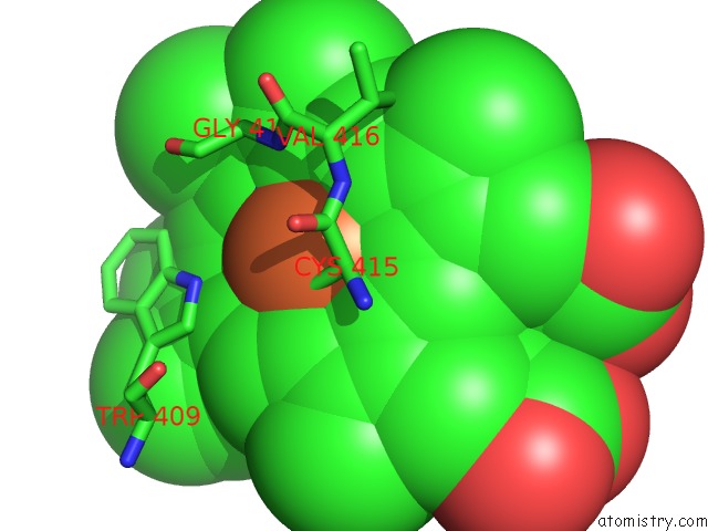
Mono view
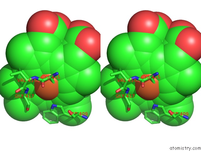
Stereo pair view
Mono view
Stereo pair view
A full contact list of Iron with other atoms in the Fe binding
site number 1 of Rat Neuronal Nitric Oxide Synthase Oxygenase Domain Complexed with Natural Substrate L-Arg. within 5.0Å range:
|
Iron binding site 2 out of 2 in 1zvl
Go back to
Iron binding site 2 out
of 2 in the Rat Neuronal Nitric Oxide Synthase Oxygenase Domain Complexed with Natural Substrate L-Arg.
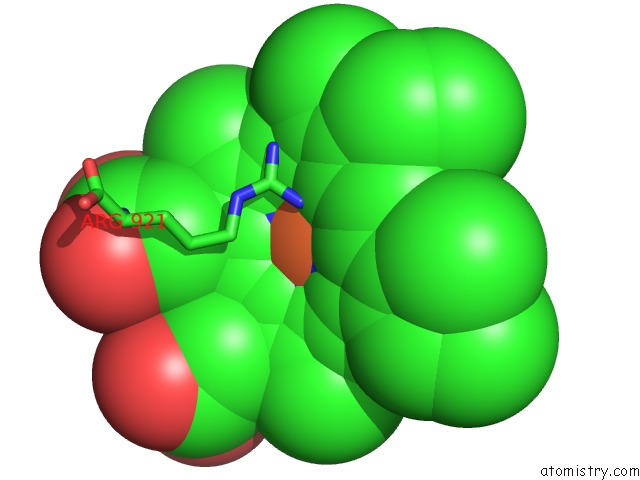
Mono view
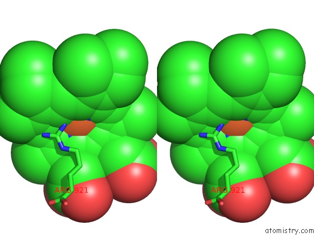
Stereo pair view
Mono view
Stereo pair view
A full contact list of Iron with other atoms in the Fe binding
site number 2 of Rat Neuronal Nitric Oxide Synthase Oxygenase Domain Complexed with Natural Substrate L-Arg. within 5.0Å range:
|
Reference:
H.Matter,
H.S.Kumar,
R.Fedorov,
A.Frey,
P.Kotsonis,
E.Hartmann,
L.G.Frohlich,
A.Reif,
W.Pfleiderer,
P.Scheurer,
D.K.Ghosh,
I.Schlichting,
H.H.Schmidt.
Structural Analysis of Isoform-Specific Inhibitors Targeting the Tetrahydrobiopterin Binding Site of Human Nitric Oxide Synthases. J.Med.Chem. V. 48 4783 2005.
ISSN: ISSN 0022-2623
PubMed: 16033258
DOI: 10.1021/JM050007X
Page generated: Sat Aug 3 18:42:32 2024
ISSN: ISSN 0022-2623
PubMed: 16033258
DOI: 10.1021/JM050007X
Last articles
Zn in 9MJ5Zn in 9HNW
Zn in 9G0L
Zn in 9FNE
Zn in 9DZN
Zn in 9E0I
Zn in 9D32
Zn in 9DAK
Zn in 8ZXC
Zn in 8ZUF