Iron »
PDB 1zlq-2ai5 »
1zz7 »
Iron in PDB 1zz7: Crystal Structure of Feii Hppe in Complex with Substrate Form 1
Protein crystallography data
The structure of Crystal Structure of Feii Hppe in Complex with Substrate Form 1, PDB code: 1zz7
was solved by
L.J.Higgins,
F.Yan,
P.Liu,
H.W.Liu,
C.L.Drennan,
with X-Ray Crystallography technique. A brief refinement statistics is given in the table below:
| Resolution Low / High (Å) | 29.03 / 2.10 |
| Space group | P 65 2 2 |
| Cell size a, b, c (Å), α, β, γ (°) | 86.560, 86.560, 220.360, 90.00, 90.00, 120.00 |
| R / Rfree (%) | 24.4 / 26.6 |
Iron Binding Sites:
The binding sites of Iron atom in the Crystal Structure of Feii Hppe in Complex with Substrate Form 1
(pdb code 1zz7). This binding sites where shown within
5.0 Angstroms radius around Iron atom.
In total 2 binding sites of Iron where determined in the Crystal Structure of Feii Hppe in Complex with Substrate Form 1, PDB code: 1zz7:
Jump to Iron binding site number: 1; 2;
In total 2 binding sites of Iron where determined in the Crystal Structure of Feii Hppe in Complex with Substrate Form 1, PDB code: 1zz7:
Jump to Iron binding site number: 1; 2;
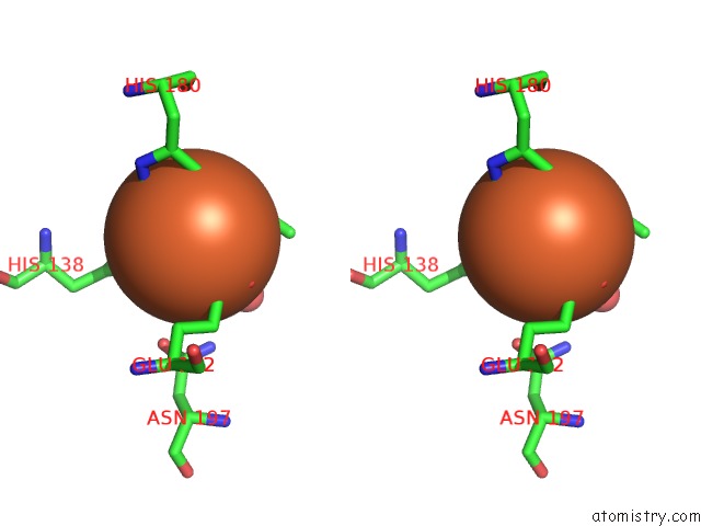
Iron binding site 1 out of 2 in 1zz7
Go back to
Iron binding site 1 out
of 2 in the Crystal Structure of Feii Hppe in Complex with Substrate Form 1
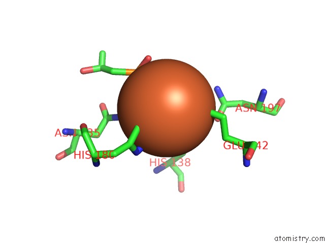
Mono view

Stereo pair view
Mono view
Stereo pair view
A full contact list of Iron with other atoms in the Fe binding
site number 1 of Crystal Structure of Feii Hppe in Complex with Substrate Form 1 within 5.0Å range:
|
Iron binding site 2 out of 2 in 1zz7
Go back to
Iron binding site 2 out
of 2 in the Crystal Structure of Feii Hppe in Complex with Substrate Form 1

Mono view
Stereo pair view
Mono view
Stereo pair view
A full contact list of Iron with other atoms in the Fe binding
site number 2 of Crystal Structure of Feii Hppe in Complex with Substrate Form 1 within 5.0Å range:
|
Reference:
L.J.Higgins,
F.Yan,
P.Liu,
H.W.Liu,
C.L.Drennan.
Structural Insight Into Antibiotic Fosfomycin Biosynthesis By A Mononuclear Iron Enzyme Nature V. 437 838 2005.
ISSN: ISSN 0028-0836
PubMed: 16015285
DOI: 10.1038/NATURE03924
Page generated: Wed Jul 16 23:20:16 2025
ISSN: ISSN 0028-0836
PubMed: 16015285
DOI: 10.1038/NATURE03924
Last articles
Fe in 2YXOFe in 2YRS
Fe in 2YXC
Fe in 2YNM
Fe in 2YVJ
Fe in 2YP1
Fe in 2YU2
Fe in 2YU1
Fe in 2YQB
Fe in 2YOO