Iron »
PDB 1zlq-2ai5 »
2a1x »
Iron in PDB 2a1x: Human Phytanoyl-Coa 2-Hydroxylase in Complex with Iron and 2- Oxoglutarate
Enzymatic activity of Human Phytanoyl-Coa 2-Hydroxylase in Complex with Iron and 2- Oxoglutarate
All present enzymatic activity of Human Phytanoyl-Coa 2-Hydroxylase in Complex with Iron and 2- Oxoglutarate:
1.14.11.18;
1.14.11.18;
Protein crystallography data
The structure of Human Phytanoyl-Coa 2-Hydroxylase in Complex with Iron and 2- Oxoglutarate, PDB code: 2a1x
was solved by
K.L.Kavanagh,
M.A.Mcdonough,
T.Searles,
D.Butler,
G.Bunkoczi,
F.Von Delft,
A.Edwards,
C.Arrowsmith,
M.Sundstrom,
C.J.Schofield,
U.Oppermann,
Structural Genomics Consortium (Sgc),
with X-Ray Crystallography technique. A brief refinement statistics is given in the table below:
| Resolution Low / High (Å) | 30.44 / 2.50 |
| Space group | I 2 2 2 |
| Cell size a, b, c (Å), α, β, γ (°) | 67.929, 86.677, 97.541, 90.00, 90.00, 90.00 |
| R / Rfree (%) | 20.4 / 27.4 |
Iron Binding Sites:
The binding sites of Iron atom in the Human Phytanoyl-Coa 2-Hydroxylase in Complex with Iron and 2- Oxoglutarate
(pdb code 2a1x). This binding sites where shown within
5.0 Angstroms radius around Iron atom.
In total only one binding site of Iron was determined in the Human Phytanoyl-Coa 2-Hydroxylase in Complex with Iron and 2- Oxoglutarate, PDB code: 2a1x:
In total only one binding site of Iron was determined in the Human Phytanoyl-Coa 2-Hydroxylase in Complex with Iron and 2- Oxoglutarate, PDB code: 2a1x:
Iron binding site 1 out of 1 in 2a1x
Go back to
Iron binding site 1 out
of 1 in the Human Phytanoyl-Coa 2-Hydroxylase in Complex with Iron and 2- Oxoglutarate
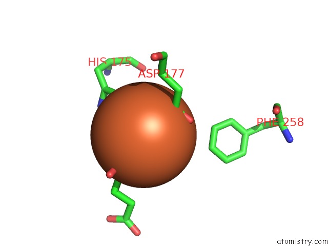
Mono view
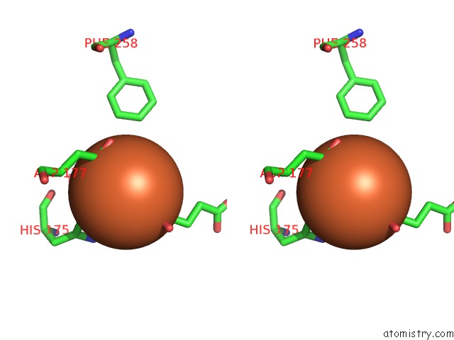
Stereo pair view
Mono view
Stereo pair view
A full contact list of Iron with other atoms in the Fe binding
site number 1 of Human Phytanoyl-Coa 2-Hydroxylase in Complex with Iron and 2- Oxoglutarate within 5.0Å range:
|
Reference:
M.A.Mcdonough,
K.L.Kavanagh,
D.Butler,
T.Searles,
U.Oppermann,
C.J.Schofield.
Structure of Human Phytanoyl-Coa 2-Hydroxylase Identifies Molecular Mechanisms of Refsum Disease J.Biol.Chem. V. 280 41101 2005.
ISSN: ISSN 0021-9258
PubMed: 16186124
DOI: 10.1074/JBC.M507528200
Page generated: Wed Jul 16 23:23:21 2025
ISSN: ISSN 0021-9258
PubMed: 16186124
DOI: 10.1074/JBC.M507528200
Last articles
Fe in 2YXOFe in 2YRS
Fe in 2YXC
Fe in 2YNM
Fe in 2YVJ
Fe in 2YP1
Fe in 2YU2
Fe in 2YU1
Fe in 2YQB
Fe in 2YOO