Iron »
PDB 1zlq-2ai5 »
2a5h »
Iron in PDB 2a5h: 2.1 Angstrom X-Ray Crystal Structure of Lysine-2,3-Aminomutase From Clostridium Subterminale SB4, with Michaelis Analog (L-Alpha-Lysine External Aldimine Form of Pyridoxal-5'-Phosphate).
Enzymatic activity of 2.1 Angstrom X-Ray Crystal Structure of Lysine-2,3-Aminomutase From Clostridium Subterminale SB4, with Michaelis Analog (L-Alpha-Lysine External Aldimine Form of Pyridoxal-5'-Phosphate).
All present enzymatic activity of 2.1 Angstrom X-Ray Crystal Structure of Lysine-2,3-Aminomutase From Clostridium Subterminale SB4, with Michaelis Analog (L-Alpha-Lysine External Aldimine Form of Pyridoxal-5'-Phosphate).:
5.4.3.2;
5.4.3.2;
Protein crystallography data
The structure of 2.1 Angstrom X-Ray Crystal Structure of Lysine-2,3-Aminomutase From Clostridium Subterminale SB4, with Michaelis Analog (L-Alpha-Lysine External Aldimine Form of Pyridoxal-5'-Phosphate)., PDB code: 2a5h
was solved by
B.W.Lepore,
F.J.Ruzicka,
P.A.Frey,
D.Ringe,
with X-Ray Crystallography technique. A brief refinement statistics is given in the table below:
| Resolution Low / High (Å) | 50.00 / 2.10 |
| Space group | C 1 2 1 |
| Cell size a, b, c (Å), α, β, γ (°) | 118.890, 92.926, 177.735, 90.00, 96.74, 90.00 |
| R / Rfree (%) | 18.4 / 22.5 |
Other elements in 2a5h:
The structure of 2.1 Angstrom X-Ray Crystal Structure of Lysine-2,3-Aminomutase From Clostridium Subterminale SB4, with Michaelis Analog (L-Alpha-Lysine External Aldimine Form of Pyridoxal-5'-Phosphate). also contains other interesting chemical elements:
| Zinc | (Zn) | 4 atoms |
Iron Binding Sites:
Pages:
>>> Page 1 <<< Page 2, Binding sites: 11 - 16;Binding sites:
The binding sites of Iron atom in the 2.1 Angstrom X-Ray Crystal Structure of Lysine-2,3-Aminomutase From Clostridium Subterminale SB4, with Michaelis Analog (L-Alpha-Lysine External Aldimine Form of Pyridoxal-5'-Phosphate). (pdb code 2a5h). This binding sites where shown within 5.0 Angstroms radius around Iron atom.In total 16 binding sites of Iron where determined in the 2.1 Angstrom X-Ray Crystal Structure of Lysine-2,3-Aminomutase From Clostridium Subterminale SB4, with Michaelis Analog (L-Alpha-Lysine External Aldimine Form of Pyridoxal-5'-Phosphate)., PDB code: 2a5h:
Jump to Iron binding site number: 1; 2; 3; 4; 5; 6; 7; 8; 9; 10;
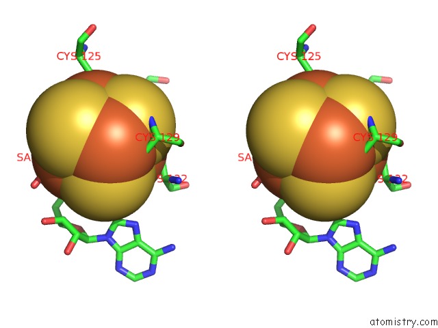
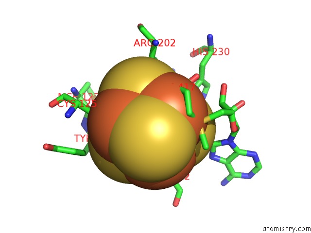
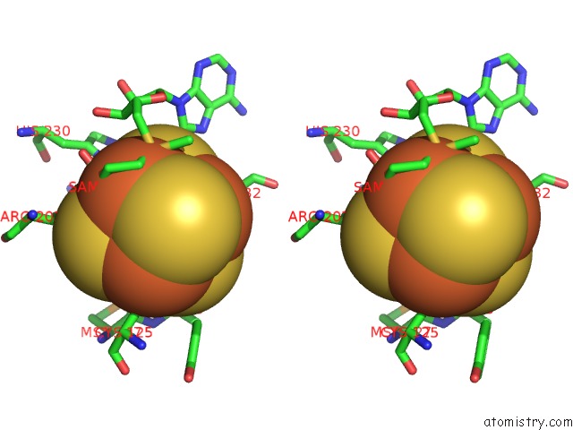
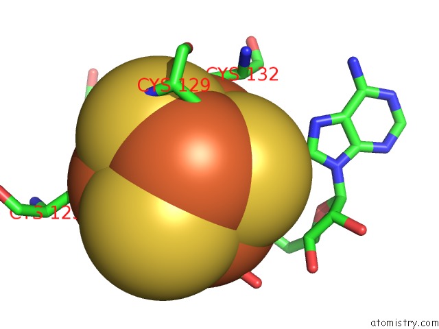
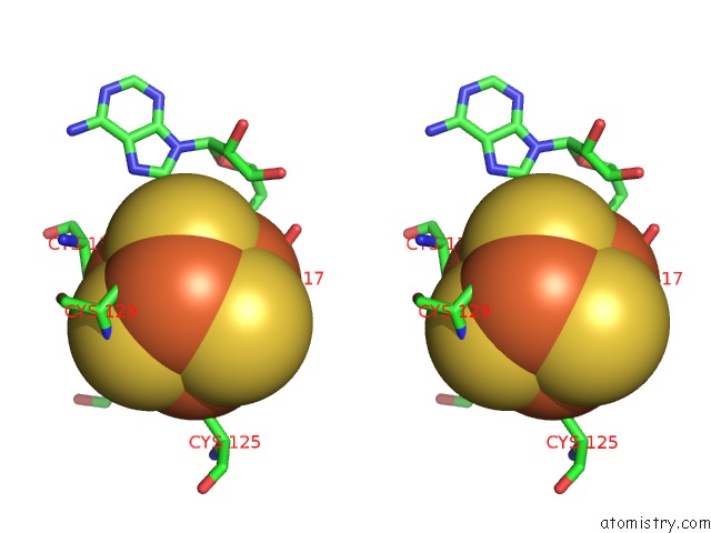
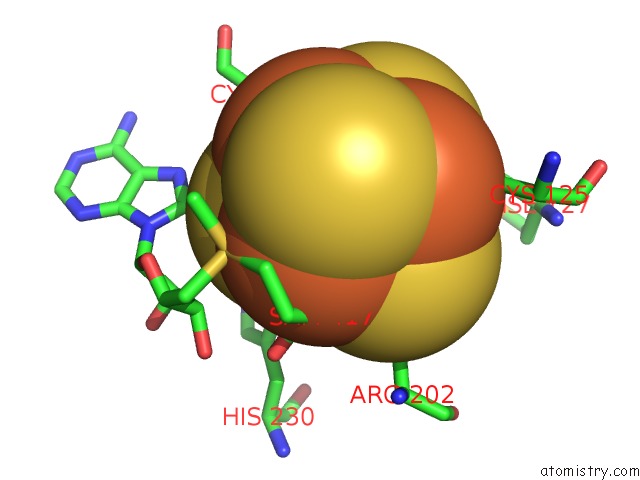
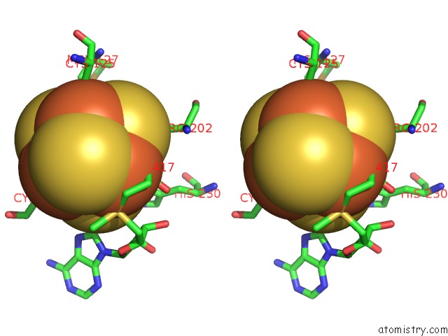
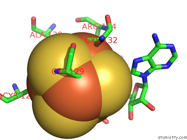
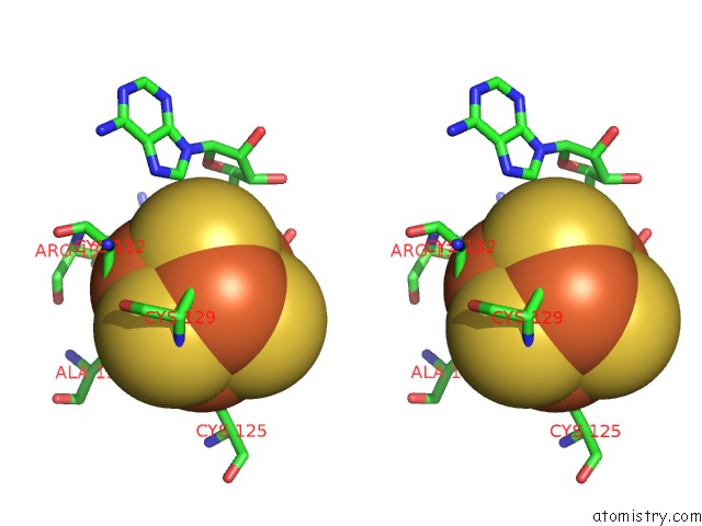
Iron binding site 1 out of 16 in 2a5h
Go back to
Iron binding site 1 out
of 16 in the 2.1 Angstrom X-Ray Crystal Structure of Lysine-2,3-Aminomutase From Clostridium Subterminale SB4, with Michaelis Analog (L-Alpha-Lysine External Aldimine Form of Pyridoxal-5'-Phosphate).

Mono view
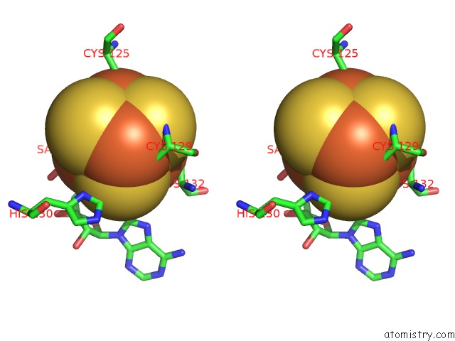
Stereo pair view
Mono view
Stereo pair view
A full contact list of Iron with other atoms in the Fe binding
site number 1 of 2.1 Angstrom X-Ray Crystal Structure of Lysine-2,3-Aminomutase From Clostridium Subterminale SB4, with Michaelis Analog (L-Alpha-Lysine External Aldimine Form of Pyridoxal-5'-Phosphate). within 5.0Å range:
|
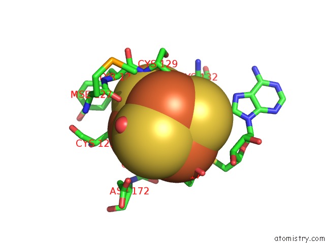
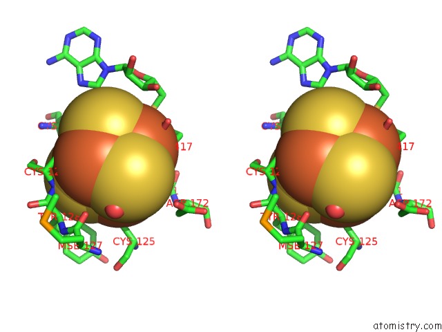
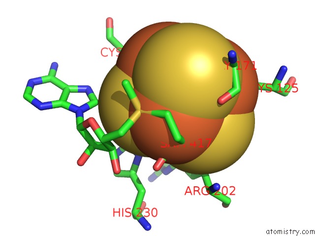
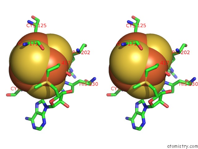
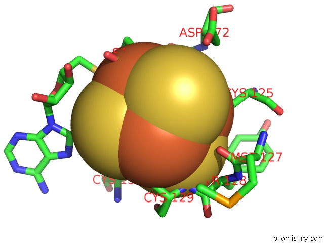
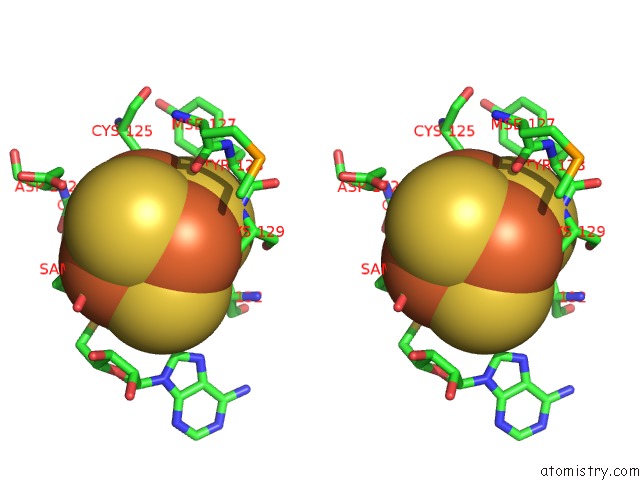
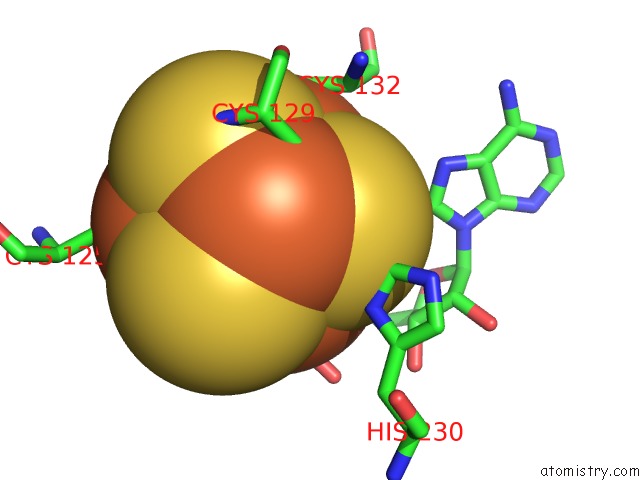
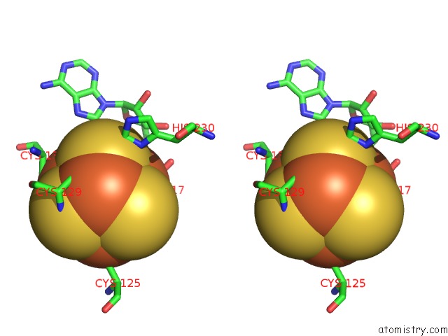
Iron binding site 2 out of 16 in 2a5h
Go back to
Iron binding site 2 out
of 16 in the 2.1 Angstrom X-Ray Crystal Structure of Lysine-2,3-Aminomutase From Clostridium Subterminale SB4, with Michaelis Analog (L-Alpha-Lysine External Aldimine Form of Pyridoxal-5'-Phosphate).

Mono view
Stereo pair view
Mono view
Stereo pair view
A full contact list of Iron with other atoms in the Fe binding
site number 2 of 2.1 Angstrom X-Ray Crystal Structure of Lysine-2,3-Aminomutase From Clostridium Subterminale SB4, with Michaelis Analog (L-Alpha-Lysine External Aldimine Form of Pyridoxal-5'-Phosphate). within 5.0Å range:
|
Iron binding site 3 out of 16 in 2a5h
Go back to
Iron binding site 3 out
of 16 in the 2.1 Angstrom X-Ray Crystal Structure of Lysine-2,3-Aminomutase From Clostridium Subterminale SB4, with Michaelis Analog (L-Alpha-Lysine External Aldimine Form of Pyridoxal-5'-Phosphate).
Mono view
Stereo pair view
Mono view
Stereo pair view
A full contact list of Iron with other atoms in the Fe binding
site number 3 of 2.1 Angstrom X-Ray Crystal Structure of Lysine-2,3-Aminomutase From Clostridium Subterminale SB4, with Michaelis Analog (L-Alpha-Lysine External Aldimine Form of Pyridoxal-5'-Phosphate). within 5.0Å range:
|
Iron binding site 4 out of 16 in 2a5h
Go back to
Iron binding site 4 out
of 16 in the 2.1 Angstrom X-Ray Crystal Structure of Lysine-2,3-Aminomutase From Clostridium Subterminale SB4, with Michaelis Analog (L-Alpha-Lysine External Aldimine Form of Pyridoxal-5'-Phosphate).
Mono view
Stereo pair view
Mono view
Stereo pair view
A full contact list of Iron with other atoms in the Fe binding
site number 4 of 2.1 Angstrom X-Ray Crystal Structure of Lysine-2,3-Aminomutase From Clostridium Subterminale SB4, with Michaelis Analog (L-Alpha-Lysine External Aldimine Form of Pyridoxal-5'-Phosphate). within 5.0Å range:
|
Iron binding site 5 out of 16 in 2a5h
Go back to
Iron binding site 5 out
of 16 in the 2.1 Angstrom X-Ray Crystal Structure of Lysine-2,3-Aminomutase From Clostridium Subterminale SB4, with Michaelis Analog (L-Alpha-Lysine External Aldimine Form of Pyridoxal-5'-Phosphate).
Mono view
Stereo pair view
Mono view
Stereo pair view
A full contact list of Iron with other atoms in the Fe binding
site number 5 of 2.1 Angstrom X-Ray Crystal Structure of Lysine-2,3-Aminomutase From Clostridium Subterminale SB4, with Michaelis Analog (L-Alpha-Lysine External Aldimine Form of Pyridoxal-5'-Phosphate). within 5.0Å range:
|
Iron binding site 6 out of 16 in 2a5h
Go back to
Iron binding site 6 out
of 16 in the 2.1 Angstrom X-Ray Crystal Structure of Lysine-2,3-Aminomutase From Clostridium Subterminale SB4, with Michaelis Analog (L-Alpha-Lysine External Aldimine Form of Pyridoxal-5'-Phosphate).
Mono view
Stereo pair view
Mono view
Stereo pair view
A full contact list of Iron with other atoms in the Fe binding
site number 6 of 2.1 Angstrom X-Ray Crystal Structure of Lysine-2,3-Aminomutase From Clostridium Subterminale SB4, with Michaelis Analog (L-Alpha-Lysine External Aldimine Form of Pyridoxal-5'-Phosphate). within 5.0Å range:
|
Iron binding site 7 out of 16 in 2a5h
Go back to
Iron binding site 7 out
of 16 in the 2.1 Angstrom X-Ray Crystal Structure of Lysine-2,3-Aminomutase From Clostridium Subterminale SB4, with Michaelis Analog (L-Alpha-Lysine External Aldimine Form of Pyridoxal-5'-Phosphate).
Mono view
Stereo pair view
Mono view
Stereo pair view
A full contact list of Iron with other atoms in the Fe binding
site number 7 of 2.1 Angstrom X-Ray Crystal Structure of Lysine-2,3-Aminomutase From Clostridium Subterminale SB4, with Michaelis Analog (L-Alpha-Lysine External Aldimine Form of Pyridoxal-5'-Phosphate). within 5.0Å range:
|
Iron binding site 8 out of 16 in 2a5h
Go back to
Iron binding site 8 out
of 16 in the 2.1 Angstrom X-Ray Crystal Structure of Lysine-2,3-Aminomutase From Clostridium Subterminale SB4, with Michaelis Analog (L-Alpha-Lysine External Aldimine Form of Pyridoxal-5'-Phosphate).
Mono view
Stereo pair view
Mono view
Stereo pair view
A full contact list of Iron with other atoms in the Fe binding
site number 8 of 2.1 Angstrom X-Ray Crystal Structure of Lysine-2,3-Aminomutase From Clostridium Subterminale SB4, with Michaelis Analog (L-Alpha-Lysine External Aldimine Form of Pyridoxal-5'-Phosphate). within 5.0Å range:
|
Iron binding site 9 out of 16 in 2a5h
Go back to
Iron binding site 9 out
of 16 in the 2.1 Angstrom X-Ray Crystal Structure of Lysine-2,3-Aminomutase From Clostridium Subterminale SB4, with Michaelis Analog (L-Alpha-Lysine External Aldimine Form of Pyridoxal-5'-Phosphate).
Mono view
Stereo pair view
Mono view
Stereo pair view
A full contact list of Iron with other atoms in the Fe binding
site number 9 of 2.1 Angstrom X-Ray Crystal Structure of Lysine-2,3-Aminomutase From Clostridium Subterminale SB4, with Michaelis Analog (L-Alpha-Lysine External Aldimine Form of Pyridoxal-5'-Phosphate). within 5.0Å range:
|
Iron binding site 10 out of 16 in 2a5h
Go back to
Iron binding site 10 out
of 16 in the 2.1 Angstrom X-Ray Crystal Structure of Lysine-2,3-Aminomutase From Clostridium Subterminale SB4, with Michaelis Analog (L-Alpha-Lysine External Aldimine Form of Pyridoxal-5'-Phosphate).
Mono view
Stereo pair view
Mono view
Stereo pair view
A full contact list of Iron with other atoms in the Fe binding
site number 10 of 2.1 Angstrom X-Ray Crystal Structure of Lysine-2,3-Aminomutase From Clostridium Subterminale SB4, with Michaelis Analog (L-Alpha-Lysine External Aldimine Form of Pyridoxal-5'-Phosphate). within 5.0Å range:
|
Reference:
B.W.Lepore,
F.J.Ruzicka,
P.A.Frey,
D.Ringe.
The X-Ray Crystal Structure of Lysine-2,3-Aminomutase From Clostridium Subterminale. Proc.Natl.Acad.Sci.Usa V. 102 13819 2005.
ISSN: ISSN 0027-8424
PubMed: 16166264
DOI: 10.1073/PNAS.0505726102
Page generated: Wed Jul 16 23:23:58 2025
ISSN: ISSN 0027-8424
PubMed: 16166264
DOI: 10.1073/PNAS.0505726102
Last articles
Fe in 2YXOFe in 2YRS
Fe in 2YXC
Fe in 2YNM
Fe in 2YVJ
Fe in 2YP1
Fe in 2YU2
Fe in 2YU1
Fe in 2YQB
Fe in 2YOO