Iron »
PDB 1zlq-2ai5 »
2abk »
Iron in PDB 2abk: Refinement of the Native Structure of Endonuclease III to A Resolution of 1.85 Angstrom
Enzymatic activity of Refinement of the Native Structure of Endonuclease III to A Resolution of 1.85 Angstrom
All present enzymatic activity of Refinement of the Native Structure of Endonuclease III to A Resolution of 1.85 Angstrom:
4.2.99.18;
4.2.99.18;
Protein crystallography data
The structure of Refinement of the Native Structure of Endonuclease III to A Resolution of 1.85 Angstrom, PDB code: 2abk
was solved by
M.M.Thayer,
J.A.Tainer,
with X-Ray Crystallography technique. A brief refinement statistics is given in the table below:
| Resolution Low / High (Å) | 10.00 / 1.85 |
| Space group | P 21 21 21 |
| Cell size a, b, c (Å), α, β, γ (°) | 48.500, 65.800, 86.800, 90.00, 90.00, 90.00 |
| R / Rfree (%) | 18.5 / 21.6 |
Iron Binding Sites:
The binding sites of Iron atom in the Refinement of the Native Structure of Endonuclease III to A Resolution of 1.85 Angstrom
(pdb code 2abk). This binding sites where shown within
5.0 Angstroms radius around Iron atom.
In total 4 binding sites of Iron where determined in the Refinement of the Native Structure of Endonuclease III to A Resolution of 1.85 Angstrom, PDB code: 2abk:
Jump to Iron binding site number: 1; 2; 3; 4;
In total 4 binding sites of Iron where determined in the Refinement of the Native Structure of Endonuclease III to A Resolution of 1.85 Angstrom, PDB code: 2abk:
Jump to Iron binding site number: 1; 2; 3; 4;
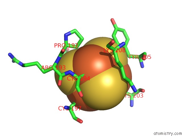
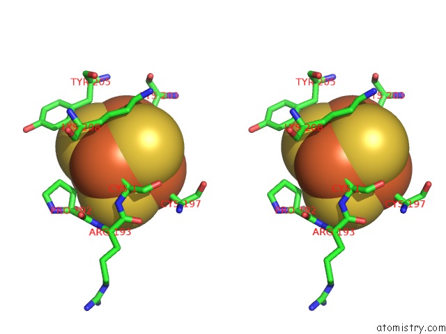
Iron binding site 1 out of 4 in 2abk
Go back to
Iron binding site 1 out
of 4 in the Refinement of the Native Structure of Endonuclease III to A Resolution of 1.85 Angstrom
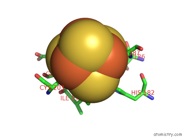
Mono view
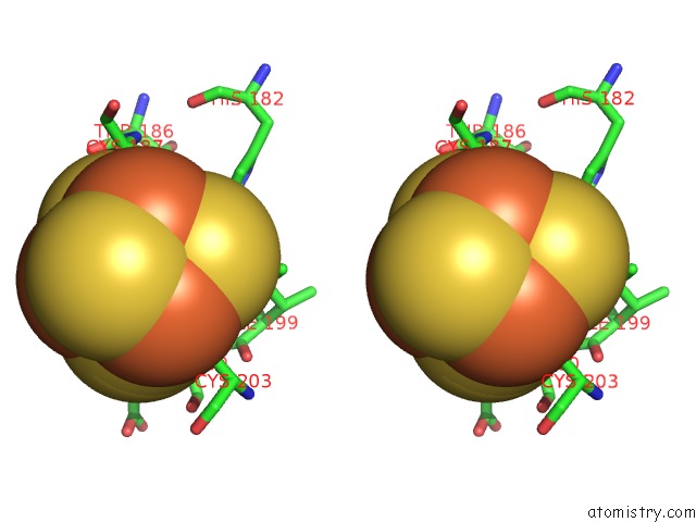
Stereo pair view
Mono view
Stereo pair view
A full contact list of Iron with other atoms in the Fe binding
site number 1 of Refinement of the Native Structure of Endonuclease III to A Resolution of 1.85 Angstrom within 5.0Å range:
|
Iron binding site 2 out of 4 in 2abk
Go back to
Iron binding site 2 out
of 4 in the Refinement of the Native Structure of Endonuclease III to A Resolution of 1.85 Angstrom
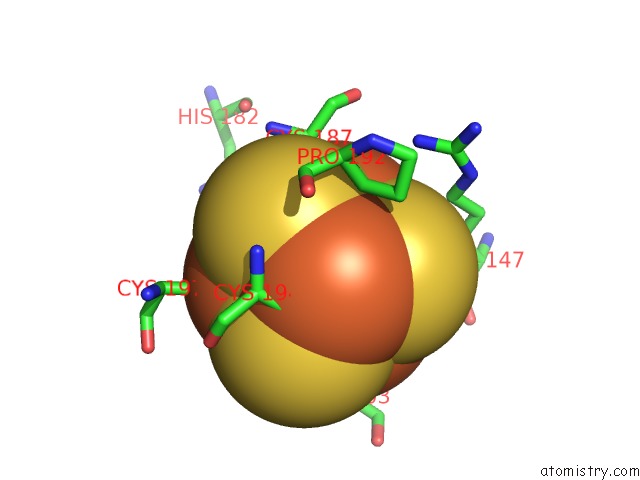
Mono view
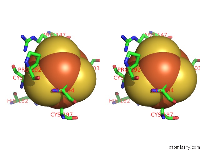
Stereo pair view
Mono view
Stereo pair view
A full contact list of Iron with other atoms in the Fe binding
site number 2 of Refinement of the Native Structure of Endonuclease III to A Resolution of 1.85 Angstrom within 5.0Å range:
|
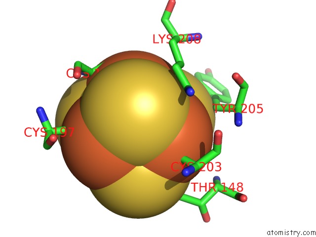
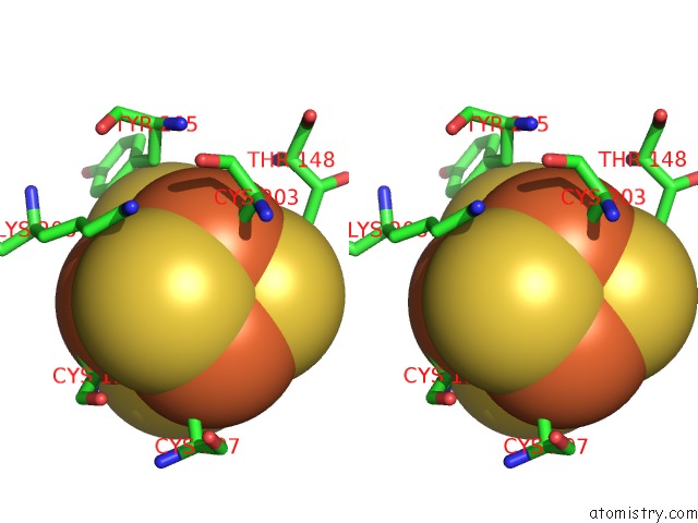
Iron binding site 3 out of 4 in 2abk
Go back to
Iron binding site 3 out
of 4 in the Refinement of the Native Structure of Endonuclease III to A Resolution of 1.85 Angstrom
Mono view
Stereo pair view
Mono view
Stereo pair view
A full contact list of Iron with other atoms in the Fe binding
site number 3 of Refinement of the Native Structure of Endonuclease III to A Resolution of 1.85 Angstrom within 5.0Å range:
|
Iron binding site 4 out of 4 in 2abk
Go back to
Iron binding site 4 out
of 4 in the Refinement of the Native Structure of Endonuclease III to A Resolution of 1.85 Angstrom
Mono view
Stereo pair view
Mono view
Stereo pair view
A full contact list of Iron with other atoms in the Fe binding
site number 4 of Refinement of the Native Structure of Endonuclease III to A Resolution of 1.85 Angstrom within 5.0Å range:
|
Reference:
M.M.Thayer,
H.Ahern,
D.Xing,
R.P.Cunningham,
J.A.Tainer.
Novel Dna Binding Motifs in the Dna Repair Enzyme Endonuclease III Crystal Structure. Embo J. V. 14 4108 1995.
ISSN: ISSN 0261-4189
PubMed: 7664751
Page generated: Sat Aug 3 18:52:54 2024
ISSN: ISSN 0261-4189
PubMed: 7664751
Last articles
Zn in 9J0NZn in 9J0O
Zn in 9J0P
Zn in 9FJX
Zn in 9EKB
Zn in 9C0F
Zn in 9CAH
Zn in 9CH0
Zn in 9CH3
Zn in 9CH1