Iron »
PDB 2ays-2bmm »
2b11 »
Iron in PDB 2b11: Crystal Structure of the Protein-Protein Complex Between F82W Cytochrome C and Cytochrome C Peroxidase
Enzymatic activity of Crystal Structure of the Protein-Protein Complex Between F82W Cytochrome C and Cytochrome C Peroxidase
All present enzymatic activity of Crystal Structure of the Protein-Protein Complex Between F82W Cytochrome C and Cytochrome C Peroxidase:
1.11.1.5;
1.11.1.5;
Protein crystallography data
The structure of Crystal Structure of the Protein-Protein Complex Between F82W Cytochrome C and Cytochrome C Peroxidase, PDB code: 2b11
was solved by
S.A.Kang,
B.R.Crane,
with X-Ray Crystallography technique. A brief refinement statistics is given in the table below:
| Resolution Low / High (Å) | 30.00 / 2.30 |
| Space group | P 1 21 1 |
| Cell size a, b, c (Å), α, β, γ (°) | 44.884, 117.864, 88.470, 90.00, 104.23, 90.00 |
| R / Rfree (%) | 26.5 / 29.3 |
Other elements in 2b11:
The structure of Crystal Structure of the Protein-Protein Complex Between F82W Cytochrome C and Cytochrome C Peroxidase also contains other interesting chemical elements:
| Zinc | (Zn) | 2 atoms |
Iron Binding Sites:
The binding sites of Iron atom in the Crystal Structure of the Protein-Protein Complex Between F82W Cytochrome C and Cytochrome C Peroxidase
(pdb code 2b11). This binding sites where shown within
5.0 Angstroms radius around Iron atom.
In total 2 binding sites of Iron where determined in the Crystal Structure of the Protein-Protein Complex Between F82W Cytochrome C and Cytochrome C Peroxidase, PDB code: 2b11:
Jump to Iron binding site number: 1; 2;
In total 2 binding sites of Iron where determined in the Crystal Structure of the Protein-Protein Complex Between F82W Cytochrome C and Cytochrome C Peroxidase, PDB code: 2b11:
Jump to Iron binding site number: 1; 2;
Iron binding site 1 out of 2 in 2b11
Go back to
Iron binding site 1 out
of 2 in the Crystal Structure of the Protein-Protein Complex Between F82W Cytochrome C and Cytochrome C Peroxidase
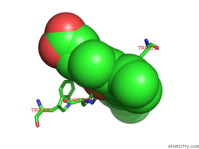
Mono view
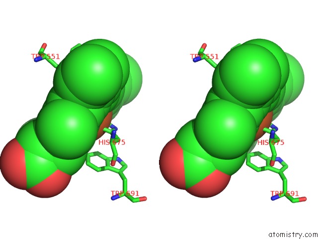
Stereo pair view
Mono view
Stereo pair view
A full contact list of Iron with other atoms in the Fe binding
site number 1 of Crystal Structure of the Protein-Protein Complex Between F82W Cytochrome C and Cytochrome C Peroxidase within 5.0Å range:
|
Iron binding site 2 out of 2 in 2b11
Go back to
Iron binding site 2 out
of 2 in the Crystal Structure of the Protein-Protein Complex Between F82W Cytochrome C and Cytochrome C Peroxidase
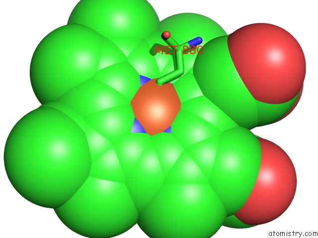
Mono view
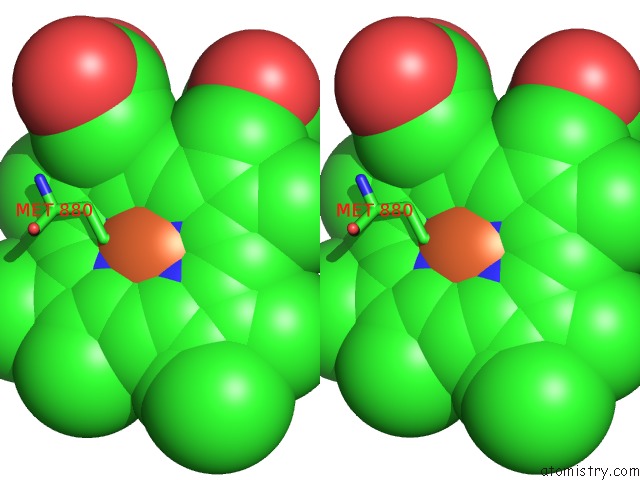
Stereo pair view
Mono view
Stereo pair view
A full contact list of Iron with other atoms in the Fe binding
site number 2 of Crystal Structure of the Protein-Protein Complex Between F82W Cytochrome C and Cytochrome C Peroxidase within 5.0Å range:
|
Reference:
S.A.Kang,
B.R.Crane.
Effects of Interface Mutations on Association Modes and Electron-Transfer Rates Between Proteins Proc.Natl.Acad.Sci.Usa V. 102 15465 2005.
ISSN: ISSN 0027-8424
PubMed: 16227441
DOI: 10.1073/PNAS.0505176102
Page generated: Sat Aug 3 19:30:42 2024
ISSN: ISSN 0027-8424
PubMed: 16227441
DOI: 10.1073/PNAS.0505176102
Last articles
Cl in 2WQLCl in 2WTA
Cl in 2WSM
Cl in 2WT8
Cl in 2WSL
Cl in 2WSA
Cl in 2WSJ
Cl in 2WS7
Cl in 2WS6
Cl in 2WQ9