Iron »
PDB 2ays-2bmm »
2b3x »
Iron in PDB 2b3x: Structure of An Orthorhombic Crystal Form of Human Cytosolic Aconitase (IRP1)
Enzymatic activity of Structure of An Orthorhombic Crystal Form of Human Cytosolic Aconitase (IRP1)
All present enzymatic activity of Structure of An Orthorhombic Crystal Form of Human Cytosolic Aconitase (IRP1):
4.2.1.3;
4.2.1.3;
Protein crystallography data
The structure of Structure of An Orthorhombic Crystal Form of Human Cytosolic Aconitase (IRP1), PDB code: 2b3x
was solved by
J.Dupuy,
J.C.Fontecilla-Camps,
A.Volbeda,
with X-Ray Crystallography technique. A brief refinement statistics is given in the table below:
| Resolution Low / High (Å) | 113.23 / 2.54 |
| Space group | C 2 2 21 |
| Cell size a, b, c (Å), α, β, γ (°) | 75.352, 103.307, 225.957, 90.00, 90.00, 90.00 |
| R / Rfree (%) | 18 / 22.5 |
Other elements in 2b3x:
The structure of Structure of An Orthorhombic Crystal Form of Human Cytosolic Aconitase (IRP1) also contains other interesting chemical elements:
| Zinc | (Zn) | 1 atom |
Iron Binding Sites:
The binding sites of Iron atom in the Structure of An Orthorhombic Crystal Form of Human Cytosolic Aconitase (IRP1)
(pdb code 2b3x). This binding sites where shown within
5.0 Angstroms radius around Iron atom.
In total 4 binding sites of Iron where determined in the Structure of An Orthorhombic Crystal Form of Human Cytosolic Aconitase (IRP1), PDB code: 2b3x:
Jump to Iron binding site number: 1; 2; 3; 4;
In total 4 binding sites of Iron where determined in the Structure of An Orthorhombic Crystal Form of Human Cytosolic Aconitase (IRP1), PDB code: 2b3x:
Jump to Iron binding site number: 1; 2; 3; 4;
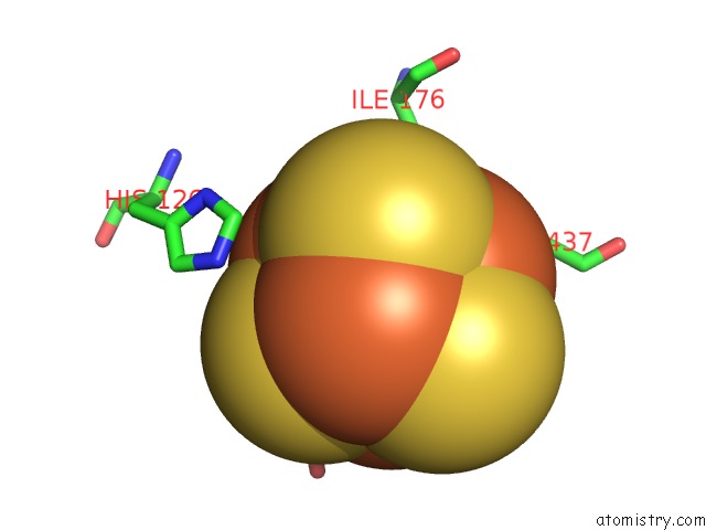
Iron binding site 1 out of 4 in 2b3x
Go back to
Iron binding site 1 out
of 4 in the Structure of An Orthorhombic Crystal Form of Human Cytosolic Aconitase (IRP1)
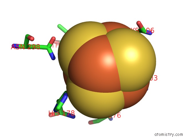
Mono view
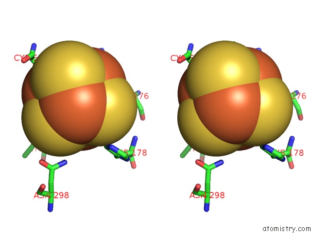
Stereo pair view
Mono view
Stereo pair view
A full contact list of Iron with other atoms in the Fe binding
site number 1 of Structure of An Orthorhombic Crystal Form of Human Cytosolic Aconitase (IRP1) within 5.0Å range:
|
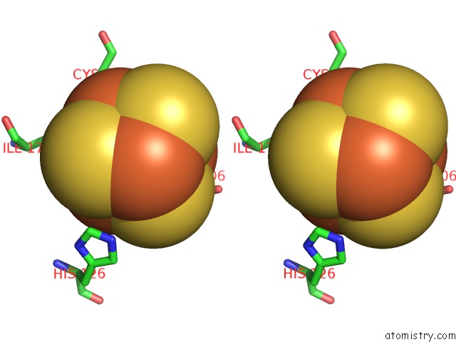
Iron binding site 2 out of 4 in 2b3x
Go back to
Iron binding site 2 out
of 4 in the Structure of An Orthorhombic Crystal Form of Human Cytosolic Aconitase (IRP1)
Mono view
Stereo pair view
Mono view
Stereo pair view
A full contact list of Iron with other atoms in the Fe binding
site number 2 of Structure of An Orthorhombic Crystal Form of Human Cytosolic Aconitase (IRP1) within 5.0Å range:
|
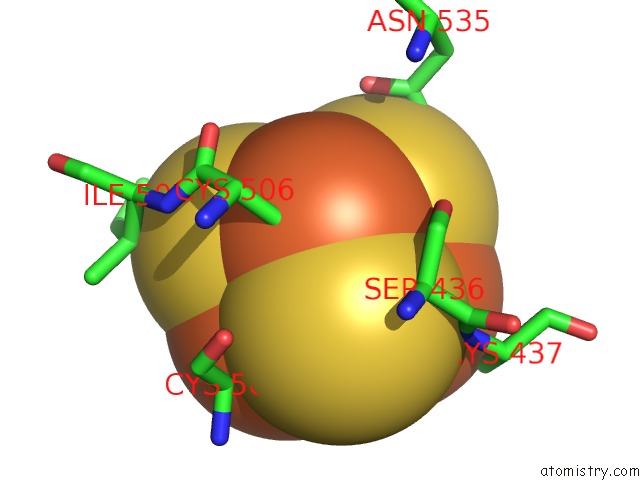
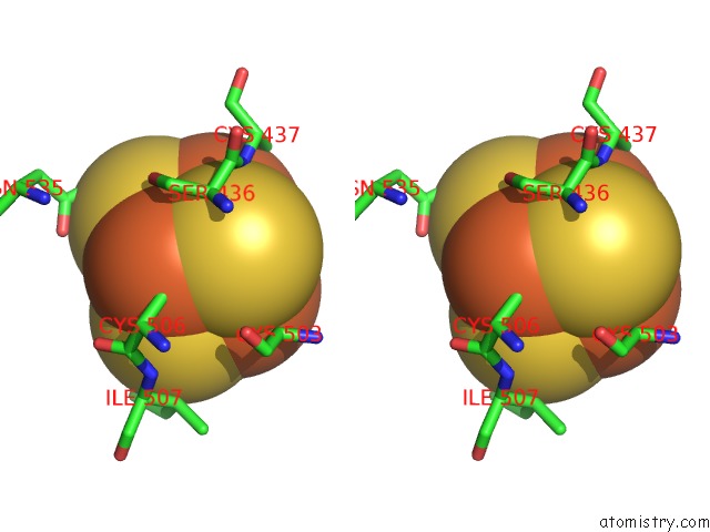
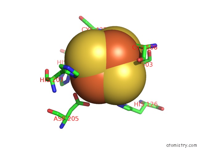
Iron binding site 3 out of 4 in 2b3x
Go back to
Iron binding site 3 out
of 4 in the Structure of An Orthorhombic Crystal Form of Human Cytosolic Aconitase (IRP1)
Mono view
Stereo pair view
Mono view
Stereo pair view
A full contact list of Iron with other atoms in the Fe binding
site number 3 of Structure of An Orthorhombic Crystal Form of Human Cytosolic Aconitase (IRP1) within 5.0Å range:
|
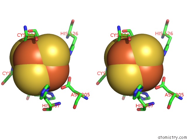
Iron binding site 4 out of 4 in 2b3x
Go back to
Iron binding site 4 out
of 4 in the Structure of An Orthorhombic Crystal Form of Human Cytosolic Aconitase (IRP1)
Mono view
Stereo pair view
Mono view
Stereo pair view
A full contact list of Iron with other atoms in the Fe binding
site number 4 of Structure of An Orthorhombic Crystal Form of Human Cytosolic Aconitase (IRP1) within 5.0Å range:
|
Reference:
J.Dupuy,
A.Volbeda,
P.Carpentier,
C.Darnault,
J.M.Moulis,
J.C.Fontecilla-Camps.
Crystal Structure of Human Iron Regulatory Protein 1 As Cytosolic Aconitase Structure V. 14 129 2006.
ISSN: ISSN 0969-2126
PubMed: 16407072
DOI: 10.1016/J.STR.2005.09.009
Page generated: Sat Aug 3 19:30:42 2024
ISSN: ISSN 0969-2126
PubMed: 16407072
DOI: 10.1016/J.STR.2005.09.009
Last articles
Zn in 9J0NZn in 9J0O
Zn in 9J0P
Zn in 9FJX
Zn in 9EKB
Zn in 9C0F
Zn in 9CAH
Zn in 9CH0
Zn in 9CH3
Zn in 9CH1