Iron »
PDB 2ays-2bmm »
2b5h »
Iron in PDB 2b5h: 1.5 A Resolution Crystal Structure of Recombinant R. Norvegicus Cysteine Dioxygenase
Enzymatic activity of 1.5 A Resolution Crystal Structure of Recombinant R. Norvegicus Cysteine Dioxygenase
All present enzymatic activity of 1.5 A Resolution Crystal Structure of Recombinant R. Norvegicus Cysteine Dioxygenase:
1.13.11.20;
1.13.11.20;
Protein crystallography data
The structure of 1.5 A Resolution Crystal Structure of Recombinant R. Norvegicus Cysteine Dioxygenase, PDB code: 2b5h
was solved by
C.R.Simmons,
Q.Liu,
Q.Huang,
Q.Hao,
T.P.Begley,
P.A.Karplus,
M.H.Stipanuk,
with X-Ray Crystallography technique. A brief refinement statistics is given in the table below:
| Resolution Low / High (Å) | 28.78 / 1.50 |
| Space group | P 43 21 2 |
| Cell size a, b, c (Å), α, β, γ (°) | 57.553, 57.553, 123.070, 90.00, 90.00, 90.00 |
| R / Rfree (%) | 18.2 / 20.8 |
Iron Binding Sites:
The binding sites of Iron atom in the 1.5 A Resolution Crystal Structure of Recombinant R. Norvegicus Cysteine Dioxygenase
(pdb code 2b5h). This binding sites where shown within
5.0 Angstroms radius around Iron atom.
In total only one binding site of Iron was determined in the 1.5 A Resolution Crystal Structure of Recombinant R. Norvegicus Cysteine Dioxygenase, PDB code: 2b5h:
In total only one binding site of Iron was determined in the 1.5 A Resolution Crystal Structure of Recombinant R. Norvegicus Cysteine Dioxygenase, PDB code: 2b5h:
Iron binding site 1 out of 1 in 2b5h
Go back to
Iron binding site 1 out
of 1 in the 1.5 A Resolution Crystal Structure of Recombinant R. Norvegicus Cysteine Dioxygenase
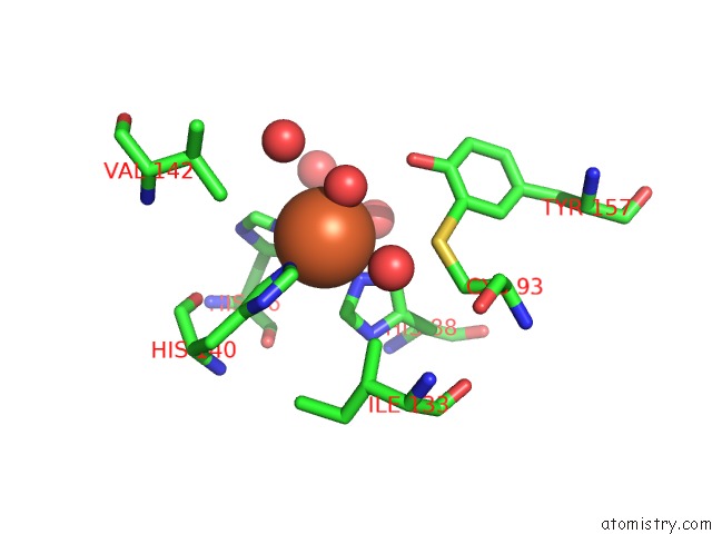
Mono view
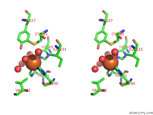
Stereo pair view
Mono view
Stereo pair view
A full contact list of Iron with other atoms in the Fe binding
site number 1 of 1.5 A Resolution Crystal Structure of Recombinant R. Norvegicus Cysteine Dioxygenase within 5.0Å range:
|
Reference:
C.R.Simmons,
Q.Liu,
Q.Huang,
Q.Hao,
T.P.Begley,
P.A.Karplus,
M.H.Stipanuk.
Crystal Structure of Mammalian Cysteine Dioxygenase: A Novel Mononuclear Iron Center For Cysteine Thiol Oxidation. J.Biol.Chem. V. 281 18723 2006.
ISSN: ISSN 0021-9258
PubMed: 16611640
DOI: 10.1074/JBC.M601555200
Page generated: Sat Aug 3 19:31:47 2024
ISSN: ISSN 0021-9258
PubMed: 16611640
DOI: 10.1074/JBC.M601555200
Last articles
Zn in 9J0NZn in 9J0O
Zn in 9J0P
Zn in 9FJX
Zn in 9EKB
Zn in 9C0F
Zn in 9CAH
Zn in 9CH0
Zn in 9CH3
Zn in 9CH1