Iron »
PDB 2ays-2bmm »
2bh2 »
Iron in PDB 2bh2: Crystal Structure of E. Coli 5-Methyluridine Methyltransferase Ruma in Complex with Ribosomal Rna Substrate and S-Adenosylhomocysteine.
Protein crystallography data
The structure of Crystal Structure of E. Coli 5-Methyluridine Methyltransferase Ruma in Complex with Ribosomal Rna Substrate and S-Adenosylhomocysteine., PDB code: 2bh2
was solved by
T.T.Lee,
S.Agarwalla,
R.M.Stroud,
with X-Ray Crystallography technique. A brief refinement statistics is given in the table below:
| Resolution Low / High (Å) | 50.00 / 2.15 |
| Space group | C 1 2 1 |
| Cell size a, b, c (Å), α, β, γ (°) | 190.061, 63.542, 112.019, 90.00, 125.15, 90.00 |
| R / Rfree (%) | 17.4 / 22.9 |
Other elements in 2bh2:
The structure of Crystal Structure of E. Coli 5-Methyluridine Methyltransferase Ruma in Complex with Ribosomal Rna Substrate and S-Adenosylhomocysteine. also contains other interesting chemical elements:
| Fluorine | (F) | 2 atoms |
Iron Binding Sites:
The binding sites of Iron atom in the Crystal Structure of E. Coli 5-Methyluridine Methyltransferase Ruma in Complex with Ribosomal Rna Substrate and S-Adenosylhomocysteine.
(pdb code 2bh2). This binding sites where shown within
5.0 Angstroms radius around Iron atom.
In total 8 binding sites of Iron where determined in the Crystal Structure of E. Coli 5-Methyluridine Methyltransferase Ruma in Complex with Ribosomal Rna Substrate and S-Adenosylhomocysteine., PDB code: 2bh2:
Jump to Iron binding site number: 1; 2; 3; 4; 5; 6; 7; 8;
In total 8 binding sites of Iron where determined in the Crystal Structure of E. Coli 5-Methyluridine Methyltransferase Ruma in Complex with Ribosomal Rna Substrate and S-Adenosylhomocysteine., PDB code: 2bh2:
Jump to Iron binding site number: 1; 2; 3; 4; 5; 6; 7; 8;
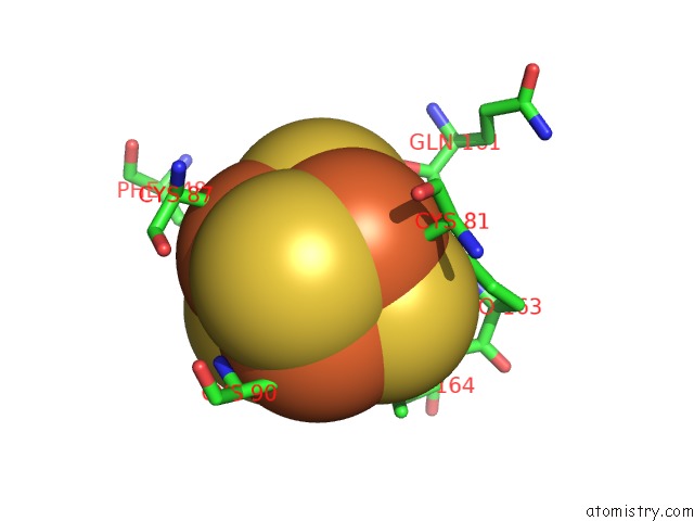
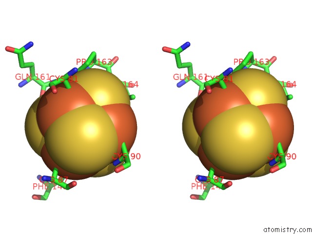
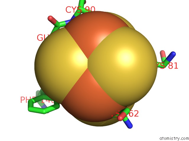
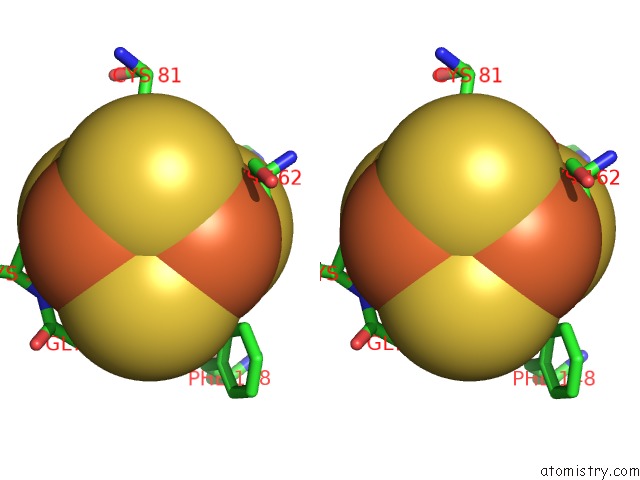
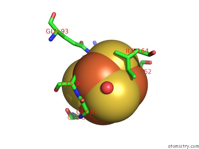
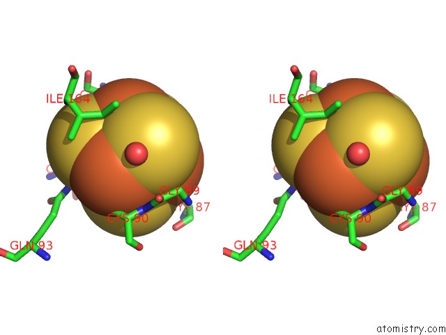
Iron binding site 1 out of 8 in 2bh2
Go back to
Iron binding site 1 out
of 8 in the Crystal Structure of E. Coli 5-Methyluridine Methyltransferase Ruma in Complex with Ribosomal Rna Substrate and S-Adenosylhomocysteine.
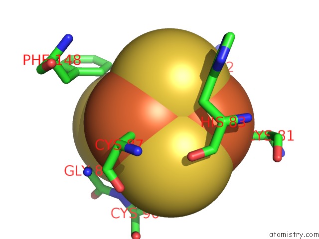
Mono view
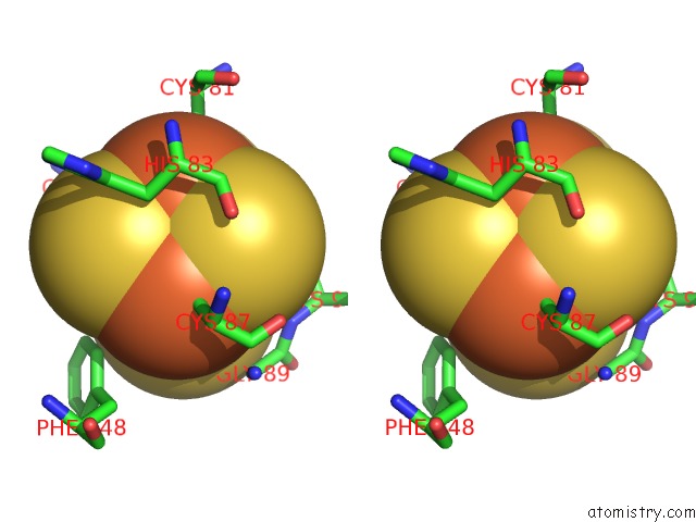
Stereo pair view
Mono view
Stereo pair view
A full contact list of Iron with other atoms in the Fe binding
site number 1 of Crystal Structure of E. Coli 5-Methyluridine Methyltransferase Ruma in Complex with Ribosomal Rna Substrate and S-Adenosylhomocysteine. within 5.0Å range:
|
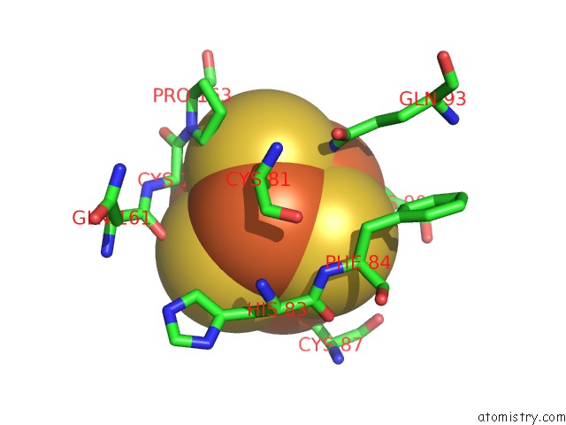
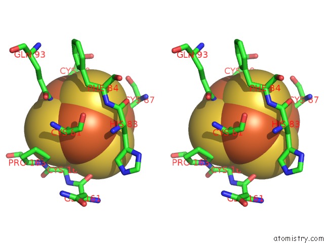
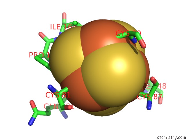
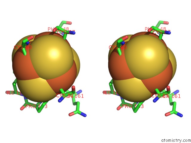
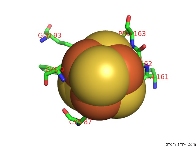
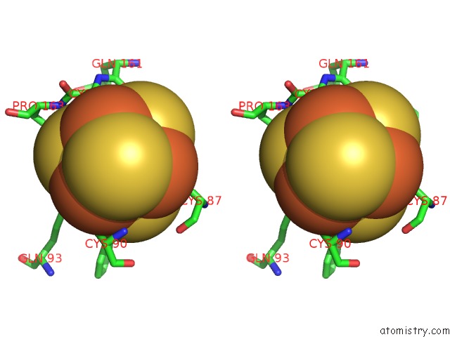
Iron binding site 2 out of 8 in 2bh2
Go back to
Iron binding site 2 out
of 8 in the Crystal Structure of E. Coli 5-Methyluridine Methyltransferase Ruma in Complex with Ribosomal Rna Substrate and S-Adenosylhomocysteine.
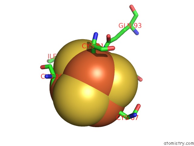
Mono view
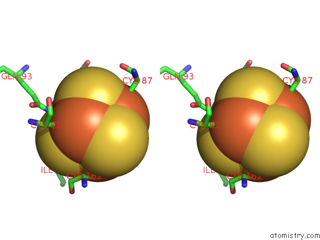
Stereo pair view
Mono view
Stereo pair view
A full contact list of Iron with other atoms in the Fe binding
site number 2 of Crystal Structure of E. Coli 5-Methyluridine Methyltransferase Ruma in Complex with Ribosomal Rna Substrate and S-Adenosylhomocysteine. within 5.0Å range:
|
Iron binding site 3 out of 8 in 2bh2
Go back to
Iron binding site 3 out
of 8 in the Crystal Structure of E. Coli 5-Methyluridine Methyltransferase Ruma in Complex with Ribosomal Rna Substrate and S-Adenosylhomocysteine.
Mono view
Stereo pair view
Mono view
Stereo pair view
A full contact list of Iron with other atoms in the Fe binding
site number 3 of Crystal Structure of E. Coli 5-Methyluridine Methyltransferase Ruma in Complex with Ribosomal Rna Substrate and S-Adenosylhomocysteine. within 5.0Å range:
|
Iron binding site 4 out of 8 in 2bh2
Go back to
Iron binding site 4 out
of 8 in the Crystal Structure of E. Coli 5-Methyluridine Methyltransferase Ruma in Complex with Ribosomal Rna Substrate and S-Adenosylhomocysteine.
Mono view
Stereo pair view
Mono view
Stereo pair view
A full contact list of Iron with other atoms in the Fe binding
site number 4 of Crystal Structure of E. Coli 5-Methyluridine Methyltransferase Ruma in Complex with Ribosomal Rna Substrate and S-Adenosylhomocysteine. within 5.0Å range:
|
Iron binding site 5 out of 8 in 2bh2
Go back to
Iron binding site 5 out
of 8 in the Crystal Structure of E. Coli 5-Methyluridine Methyltransferase Ruma in Complex with Ribosomal Rna Substrate and S-Adenosylhomocysteine.
Mono view
Stereo pair view
Mono view
Stereo pair view
A full contact list of Iron with other atoms in the Fe binding
site number 5 of Crystal Structure of E. Coli 5-Methyluridine Methyltransferase Ruma in Complex with Ribosomal Rna Substrate and S-Adenosylhomocysteine. within 5.0Å range:
|
Iron binding site 6 out of 8 in 2bh2
Go back to
Iron binding site 6 out
of 8 in the Crystal Structure of E. Coli 5-Methyluridine Methyltransferase Ruma in Complex with Ribosomal Rna Substrate and S-Adenosylhomocysteine.
Mono view
Stereo pair view
Mono view
Stereo pair view
A full contact list of Iron with other atoms in the Fe binding
site number 6 of Crystal Structure of E. Coli 5-Methyluridine Methyltransferase Ruma in Complex with Ribosomal Rna Substrate and S-Adenosylhomocysteine. within 5.0Å range:
|
Iron binding site 7 out of 8 in 2bh2
Go back to
Iron binding site 7 out
of 8 in the Crystal Structure of E. Coli 5-Methyluridine Methyltransferase Ruma in Complex with Ribosomal Rna Substrate and S-Adenosylhomocysteine.
Mono view
Stereo pair view
Mono view
Stereo pair view
A full contact list of Iron with other atoms in the Fe binding
site number 7 of Crystal Structure of E. Coli 5-Methyluridine Methyltransferase Ruma in Complex with Ribosomal Rna Substrate and S-Adenosylhomocysteine. within 5.0Å range:
|
Iron binding site 8 out of 8 in 2bh2
Go back to
Iron binding site 8 out
of 8 in the Crystal Structure of E. Coli 5-Methyluridine Methyltransferase Ruma in Complex with Ribosomal Rna Substrate and S-Adenosylhomocysteine.
Mono view
Stereo pair view
Mono view
Stereo pair view
A full contact list of Iron with other atoms in the Fe binding
site number 8 of Crystal Structure of E. Coli 5-Methyluridine Methyltransferase Ruma in Complex with Ribosomal Rna Substrate and S-Adenosylhomocysteine. within 5.0Å range:
|
Reference:
T.T.Lee,
S.Agarwalla,
R.M.Stroud.
A Unique Rna Fold in the Ruma-Rna-Cofactor Ternary Complex Contributes to Substrate Selectivity and Enzymatic Function Cell(Cambridge,Mass.) V. 120 599 2005.
ISSN: ISSN 0092-8674
PubMed: 15766524
DOI: 10.1016/J.CELL.2004.12.037
Page generated: Sat Aug 3 19:36:52 2024
ISSN: ISSN 0092-8674
PubMed: 15766524
DOI: 10.1016/J.CELL.2004.12.037
Last articles
Zn in 9J0NZn in 9J0O
Zn in 9J0P
Zn in 9FJX
Zn in 9EKB
Zn in 9C0F
Zn in 9CAH
Zn in 9CH0
Zn in 9CH3
Zn in 9CH1