Iron »
PDB 2ays-2bmm »
2blj »
Iron in PDB 2blj: Structure of L29W Mbco
Protein crystallography data
The structure of Structure of L29W Mbco, PDB code: 2blj
was solved by
K.Nienhaus,
A.Ostermann,
G.U.Nienhaus,
F.G.Parak,
M.Schmidt,
with X-Ray Crystallography technique. A brief refinement statistics is given in the table below:
| Resolution Low / High (Å) | 50.00 / 1.80 |
| Space group | P 6 |
| Cell size a, b, c (Å), α, β, γ (°) | 91.870, 91.870, 46.040, 90.00, 90.00, 120.00 |
| R / Rfree (%) | 22.4 / 25.7 |
Iron Binding Sites:
The binding sites of Iron atom in the Structure of L29W Mbco
(pdb code 2blj). This binding sites where shown within
5.0 Angstroms radius around Iron atom.
In total only one binding site of Iron was determined in the Structure of L29W Mbco, PDB code: 2blj:
In total only one binding site of Iron was determined in the Structure of L29W Mbco, PDB code: 2blj:
Iron binding site 1 out of 1 in 2blj
Go back to
Iron binding site 1 out
of 1 in the Structure of L29W Mbco
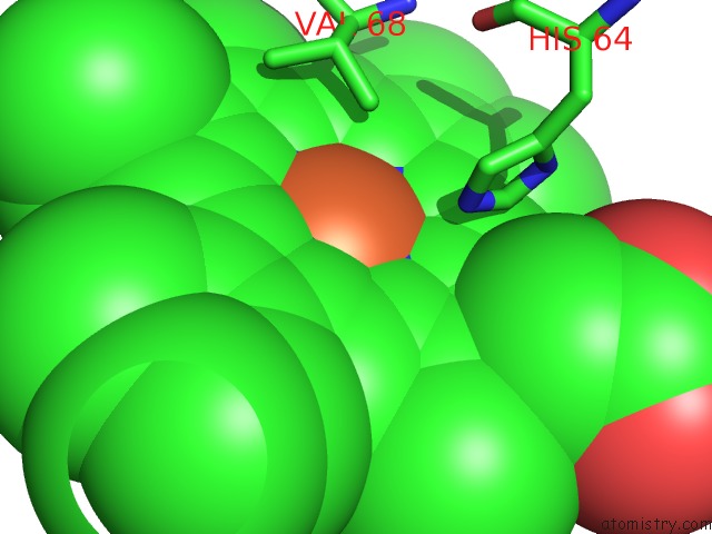
Mono view
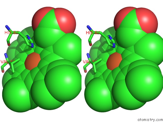
Stereo pair view
Mono view
Stereo pair view
A full contact list of Iron with other atoms in the Fe binding
site number 1 of Structure of L29W Mbco within 5.0Å range:
|
Reference:
K.Nienhaus,
A.Ostermann,
G.U.Nienhaus,
F.G.Parak,
M.Schmidt.
Ligand Migration and Protein Fluctuations in Myoglobin Mutant L29W Biochemistry V. 44 5095 2005.
ISSN: ISSN 0006-2960
PubMed: 15794647
DOI: 10.1021/BI047513T
Page generated: Sat Aug 3 19:40:50 2024
ISSN: ISSN 0006-2960
PubMed: 15794647
DOI: 10.1021/BI047513T
Last articles
Zn in 9MJ5Zn in 9HNW
Zn in 9G0L
Zn in 9FNE
Zn in 9DZN
Zn in 9E0I
Zn in 9D32
Zn in 9DAK
Zn in 8ZXC
Zn in 8ZUF