Iron »
PDB 2bmo-2c1v »
2bq4 »
Iron in PDB 2bq4: Crystal Structure of Type I Cytochrome C3 From Desulfovibrio Africanus
Protein crystallography data
The structure of Crystal Structure of Type I Cytochrome C3 From Desulfovibrio Africanus, PDB code: 2bq4
was solved by
M.Czjzek,
L.Pieulle,
X.Morelli,
F.Guerlesquin,
E.C.Hatchikian,
with X-Ray Crystallography technique. A brief refinement statistics is given in the table below:
| Resolution Low / High (Å) | 48.22 / 1.68 |
| Space group | C 1 2 1 |
| Cell size a, b, c (Å), α, β, γ (°) | 100.900, 46.300, 54.816, 90.00, 118.97, 90.00 |
| R / Rfree (%) | 20 / 24 |
Other elements in 2bq4:
The structure of Crystal Structure of Type I Cytochrome C3 From Desulfovibrio Africanus also contains other interesting chemical elements:
| Calcium | (Ca) | 2 atoms |
Iron Binding Sites:
The binding sites of Iron atom in the Crystal Structure of Type I Cytochrome C3 From Desulfovibrio Africanus
(pdb code 2bq4). This binding sites where shown within
5.0 Angstroms radius around Iron atom.
In total 8 binding sites of Iron where determined in the Crystal Structure of Type I Cytochrome C3 From Desulfovibrio Africanus, PDB code: 2bq4:
Jump to Iron binding site number: 1; 2; 3; 4; 5; 6; 7; 8;
In total 8 binding sites of Iron where determined in the Crystal Structure of Type I Cytochrome C3 From Desulfovibrio Africanus, PDB code: 2bq4:
Jump to Iron binding site number: 1; 2; 3; 4; 5; 6; 7; 8;
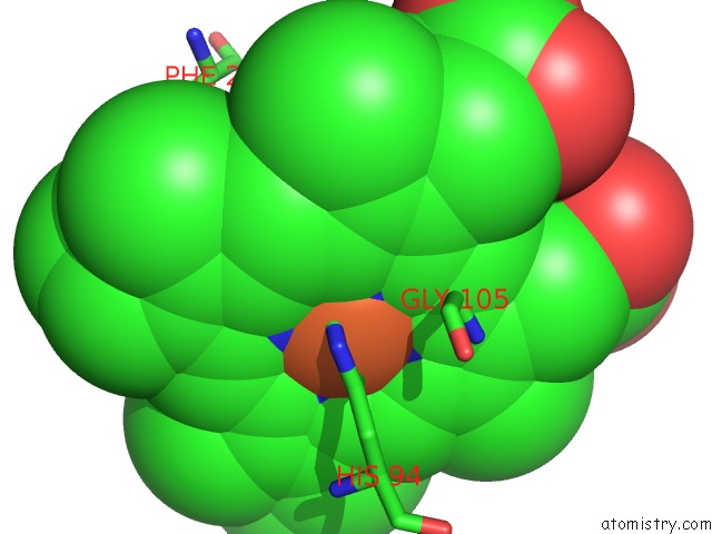
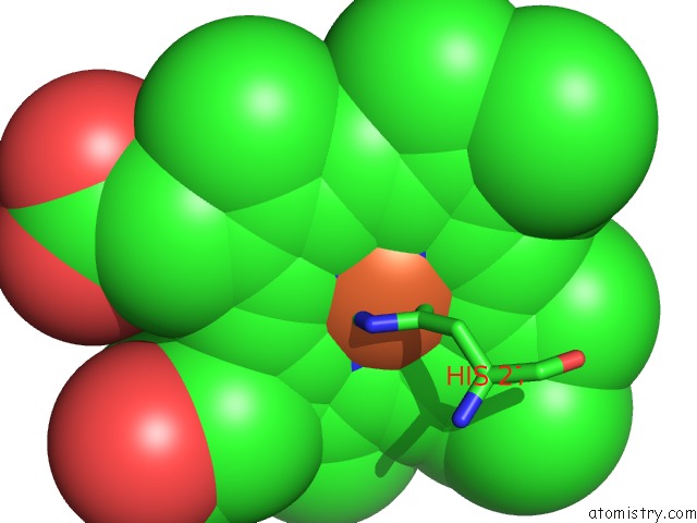
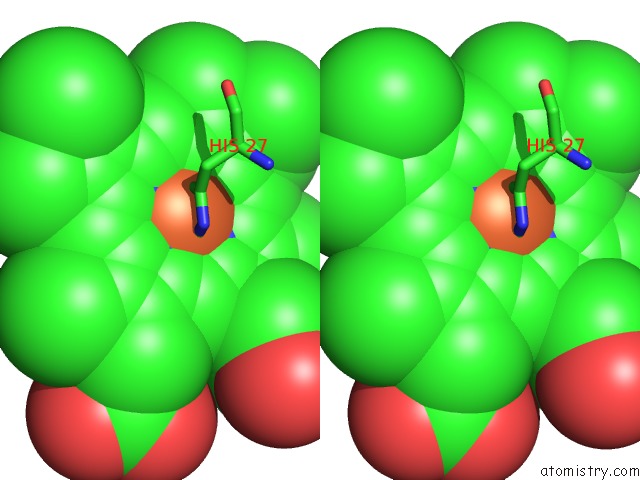
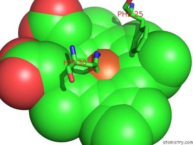
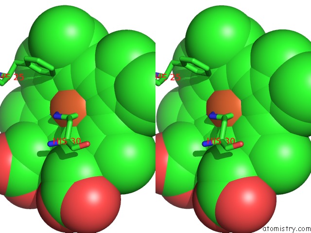
Iron binding site 1 out of 8 in 2bq4
Go back to
Iron binding site 1 out
of 8 in the Crystal Structure of Type I Cytochrome C3 From Desulfovibrio Africanus
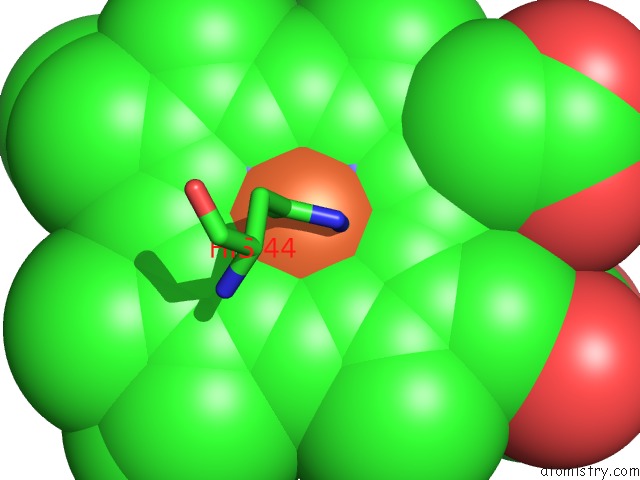
Mono view
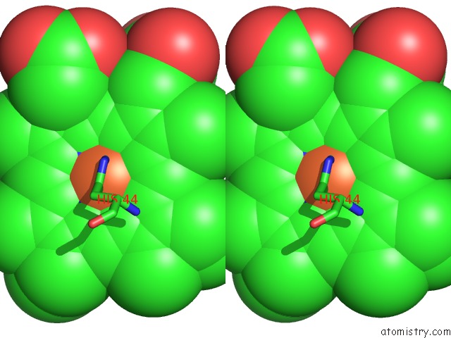
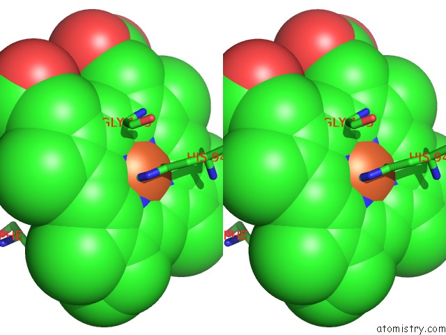
Stereo pair view
Mono view
Stereo pair view
A full contact list of Iron with other atoms in the Fe binding
site number 1 of Crystal Structure of Type I Cytochrome C3 From Desulfovibrio Africanus within 5.0Å range:
|
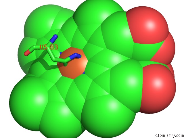
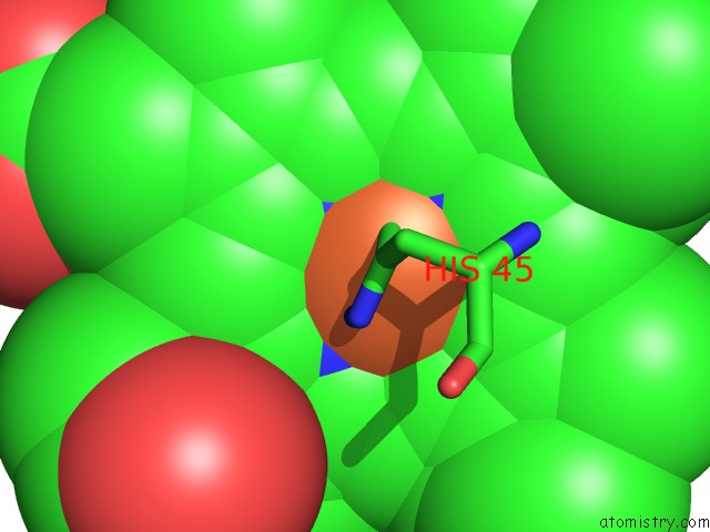
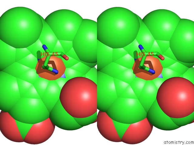
Iron binding site 2 out of 8 in 2bq4
Go back to
Iron binding site 2 out
of 8 in the Crystal Structure of Type I Cytochrome C3 From Desulfovibrio Africanus
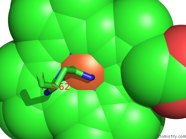
Mono view
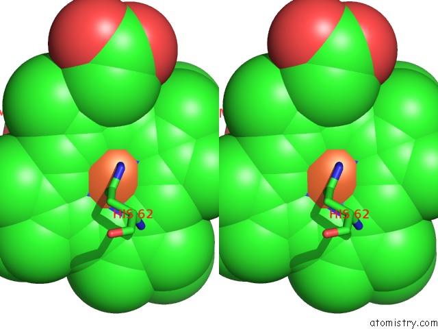
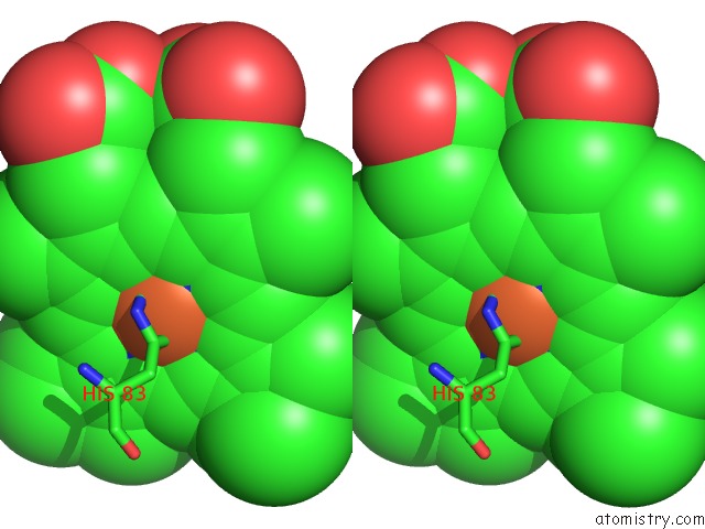
Stereo pair view
Mono view
Stereo pair view
A full contact list of Iron with other atoms in the Fe binding
site number 2 of Crystal Structure of Type I Cytochrome C3 From Desulfovibrio Africanus within 5.0Å range:
|
Iron binding site 3 out of 8 in 2bq4
Go back to
Iron binding site 3 out
of 8 in the Crystal Structure of Type I Cytochrome C3 From Desulfovibrio Africanus
Mono view
Stereo pair view
Mono view
Stereo pair view
A full contact list of Iron with other atoms in the Fe binding
site number 3 of Crystal Structure of Type I Cytochrome C3 From Desulfovibrio Africanus within 5.0Å range:
|
Iron binding site 4 out of 8 in 2bq4
Go back to
Iron binding site 4 out
of 8 in the Crystal Structure of Type I Cytochrome C3 From Desulfovibrio Africanus
Mono view
Stereo pair view
Mono view
Stereo pair view
A full contact list of Iron with other atoms in the Fe binding
site number 4 of Crystal Structure of Type I Cytochrome C3 From Desulfovibrio Africanus within 5.0Å range:
|
Iron binding site 5 out of 8 in 2bq4
Go back to
Iron binding site 5 out
of 8 in the Crystal Structure of Type I Cytochrome C3 From Desulfovibrio Africanus
Mono view
Stereo pair view
Mono view
Stereo pair view
A full contact list of Iron with other atoms in the Fe binding
site number 5 of Crystal Structure of Type I Cytochrome C3 From Desulfovibrio Africanus within 5.0Å range:
|
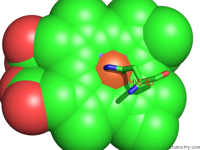
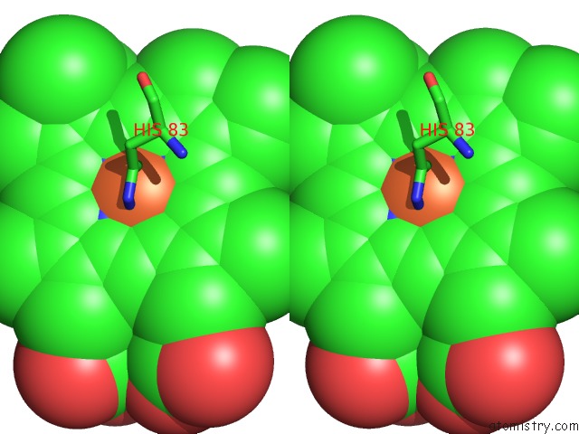
Iron binding site 6 out of 8 in 2bq4
Go back to
Iron binding site 6 out
of 8 in the Crystal Structure of Type I Cytochrome C3 From Desulfovibrio Africanus
Mono view
Stereo pair view
Mono view
Stereo pair view
A full contact list of Iron with other atoms in the Fe binding
site number 6 of Crystal Structure of Type I Cytochrome C3 From Desulfovibrio Africanus within 5.0Å range:
|
Iron binding site 7 out of 8 in 2bq4
Go back to
Iron binding site 7 out
of 8 in the Crystal Structure of Type I Cytochrome C3 From Desulfovibrio Africanus
Mono view
Stereo pair view
Mono view
Stereo pair view
A full contact list of Iron with other atoms in the Fe binding
site number 7 of Crystal Structure of Type I Cytochrome C3 From Desulfovibrio Africanus within 5.0Å range:
|
Iron binding site 8 out of 8 in 2bq4
Go back to
Iron binding site 8 out
of 8 in the Crystal Structure of Type I Cytochrome C3 From Desulfovibrio Africanus
Mono view
Stereo pair view
Mono view
Stereo pair view
A full contact list of Iron with other atoms in the Fe binding
site number 8 of Crystal Structure of Type I Cytochrome C3 From Desulfovibrio Africanus within 5.0Å range:
|
Reference:
L.Pieulle,
X.Morelli,
P.Gallice,
E.Lojou,
P.Barbier,
M.Czjzek,
P.Bianco,
F.Guerlesquin,
E.C.Hatchikian.
The Type I / Type II Cytochrome C(3) Complex: An Electron Transfer Link in the Hydrogen-Sulfate Reduction Pathway. J.Mol.Biol. V. 354 73 2005.
ISSN: ISSN 0022-2836
PubMed: 16226767
DOI: 10.1016/J.JMB.2005.09.036
Page generated: Sat Aug 3 19:46:13 2024
ISSN: ISSN 0022-2836
PubMed: 16226767
DOI: 10.1016/J.JMB.2005.09.036
Last articles
Cl in 3B01Cl in 3AZN
Cl in 3B0G
Cl in 3AZM
Cl in 3AZL
Cl in 3AZK
Cl in 3AZJ
Cl in 3AZI
Cl in 3AZ9
Cl in 3AZG