Iron »
PDB 2bmo-2c1v »
2bwh »
Iron in PDB 2bwh: Laue Structure of A Short Lived State of L29W Myoglobin
Protein crystallography data
The structure of Laue Structure of A Short Lived State of L29W Myoglobin, PDB code: 2bwh
was solved by
M.Schmidt,
K.Nienhaus,
R.Pahl,
A.Krasselt,
S.Anderson,
F.Parak,
G.U.Nienhaus,
V.Srajer,
with X-Ray Crystallography technique. A brief refinement statistics is given in the table below:
| Resolution Low / High (Å) | 15.00 / 1.90 |
| Space group | P 6 |
| Cell size a, b, c (Å), α, β, γ (°) | 91.870, 91.870, 46.040, 90.00, 90.00, 120.00 |
| R / Rfree (%) | 23 / 23.3 |
Iron Binding Sites:
The binding sites of Iron atom in the Laue Structure of A Short Lived State of L29W Myoglobin
(pdb code 2bwh). This binding sites where shown within
5.0 Angstroms radius around Iron atom.
In total only one binding site of Iron was determined in the Laue Structure of A Short Lived State of L29W Myoglobin, PDB code: 2bwh:
In total only one binding site of Iron was determined in the Laue Structure of A Short Lived State of L29W Myoglobin, PDB code: 2bwh:
Iron binding site 1 out of 1 in 2bwh
Go back to
Iron binding site 1 out
of 1 in the Laue Structure of A Short Lived State of L29W Myoglobin
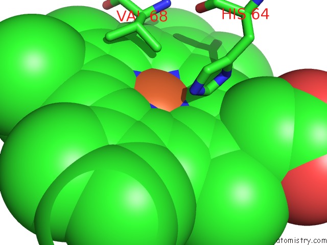
Mono view
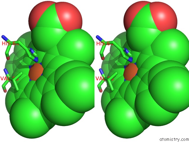
Stereo pair view
Mono view
Stereo pair view
A full contact list of Iron with other atoms in the Fe binding
site number 1 of Laue Structure of A Short Lived State of L29W Myoglobin within 5.0Å range:
|
Reference:
M.Schmidt,
K.Nienhaus,
R.Pahl,
A.Krasselt,
S.Anderson,
F.Parak,
G.U.Nienhaus,
V.Srajer.
Ligand Migration Pathway and Protein Dynamics in Myoglobin: A Time-Resolved Crystallographic Study on L29W Mbco. Proc. Natl. Acad. Sci. V. 102 11704 2005U.S.A..
ISSN: ISSN 0027-8424
PubMed: 16085709
DOI: 10.1073/PNAS.0504932102
Page generated: Thu Jul 17 00:07:07 2025
ISSN: ISSN 0027-8424
PubMed: 16085709
DOI: 10.1073/PNAS.0504932102
Last articles
Fe in 2YXOFe in 2YRS
Fe in 2YXC
Fe in 2YNM
Fe in 2YVJ
Fe in 2YP1
Fe in 2YU2
Fe in 2YU1
Fe in 2YQB
Fe in 2YOO