Iron »
PDB 2bmo-2c1v »
2c1v »
Iron in PDB 2c1v: Crystal Structure of the Di-Haem Cytochrome C Peroxidase From Paracoccus Pantotrophus - Mixed Valence Form
Enzymatic activity of Crystal Structure of the Di-Haem Cytochrome C Peroxidase From Paracoccus Pantotrophus - Mixed Valence Form
All present enzymatic activity of Crystal Structure of the Di-Haem Cytochrome C Peroxidase From Paracoccus Pantotrophus - Mixed Valence Form:
1.11.1.5;
1.11.1.5;
Protein crystallography data
The structure of Crystal Structure of the Di-Haem Cytochrome C Peroxidase From Paracoccus Pantotrophus - Mixed Valence Form, PDB code: 2c1v
was solved by
A.Echalier,
V.Fulop,
with X-Ray Crystallography technique. A brief refinement statistics is given in the table below:
| Resolution Low / High (Å) | 48.22 / 1.2 |
| Space group | P 21 21 2 |
| Cell size a, b, c (Å), α, β, γ (°) | 101.370, 141.200, 51.580, 90.00, 90.00, 90.00 |
| R / Rfree (%) | 17.3 / 18.8 |
Other elements in 2c1v:
The structure of Crystal Structure of the Di-Haem Cytochrome C Peroxidase From Paracoccus Pantotrophus - Mixed Valence Form also contains other interesting chemical elements:
| Calcium | (Ca) | 2 atoms |
Iron Binding Sites:
The binding sites of Iron atom in the Crystal Structure of the Di-Haem Cytochrome C Peroxidase From Paracoccus Pantotrophus - Mixed Valence Form
(pdb code 2c1v). This binding sites where shown within
5.0 Angstroms radius around Iron atom.
In total 4 binding sites of Iron where determined in the Crystal Structure of the Di-Haem Cytochrome C Peroxidase From Paracoccus Pantotrophus - Mixed Valence Form, PDB code: 2c1v:
Jump to Iron binding site number: 1; 2; 3; 4;
In total 4 binding sites of Iron where determined in the Crystal Structure of the Di-Haem Cytochrome C Peroxidase From Paracoccus Pantotrophus - Mixed Valence Form, PDB code: 2c1v:
Jump to Iron binding site number: 1; 2; 3; 4;
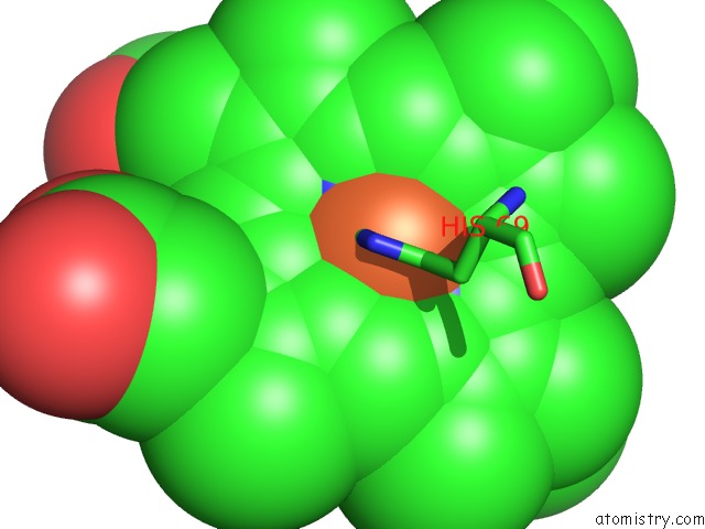
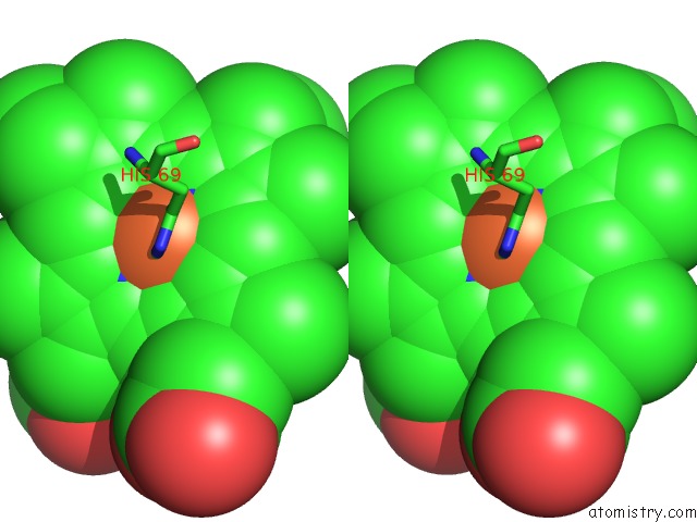
Iron binding site 1 out of 4 in 2c1v
Go back to
Iron binding site 1 out
of 4 in the Crystal Structure of the Di-Haem Cytochrome C Peroxidase From Paracoccus Pantotrophus - Mixed Valence Form
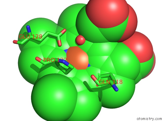
Mono view
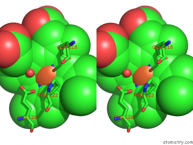
Stereo pair view
Mono view
Stereo pair view
A full contact list of Iron with other atoms in the Fe binding
site number 1 of Crystal Structure of the Di-Haem Cytochrome C Peroxidase From Paracoccus Pantotrophus - Mixed Valence Form within 5.0Å range:
|
Iron binding site 2 out of 4 in 2c1v
Go back to
Iron binding site 2 out
of 4 in the Crystal Structure of the Di-Haem Cytochrome C Peroxidase From Paracoccus Pantotrophus - Mixed Valence Form
Mono view
Stereo pair view
Mono view
Stereo pair view
A full contact list of Iron with other atoms in the Fe binding
site number 2 of Crystal Structure of the Di-Haem Cytochrome C Peroxidase From Paracoccus Pantotrophus - Mixed Valence Form within 5.0Å range:
|
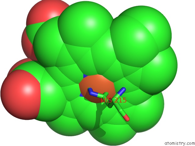
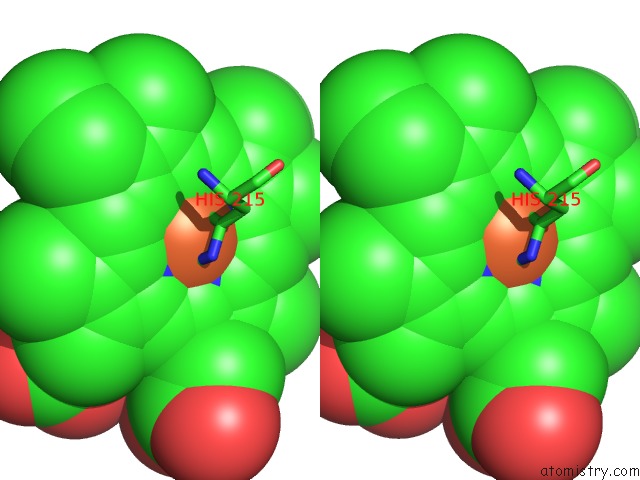
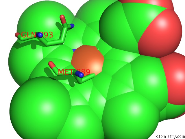
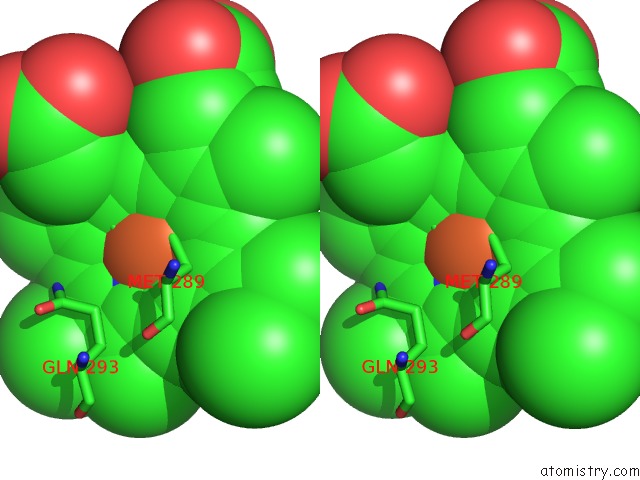
Iron binding site 3 out of 4 in 2c1v
Go back to
Iron binding site 3 out
of 4 in the Crystal Structure of the Di-Haem Cytochrome C Peroxidase From Paracoccus Pantotrophus - Mixed Valence Form
Mono view
Stereo pair view
Mono view
Stereo pair view
A full contact list of Iron with other atoms in the Fe binding
site number 3 of Crystal Structure of the Di-Haem Cytochrome C Peroxidase From Paracoccus Pantotrophus - Mixed Valence Form within 5.0Å range:
|
Iron binding site 4 out of 4 in 2c1v
Go back to
Iron binding site 4 out
of 4 in the Crystal Structure of the Di-Haem Cytochrome C Peroxidase From Paracoccus Pantotrophus - Mixed Valence Form
Mono view
Stereo pair view
Mono view
Stereo pair view
A full contact list of Iron with other atoms in the Fe binding
site number 4 of Crystal Structure of the Di-Haem Cytochrome C Peroxidase From Paracoccus Pantotrophus - Mixed Valence Form within 5.0Å range:
|
Reference:
A.Echalier,
C.F.Goodhew,
G.W.Pettigrew,
V.Fulop.
Activation and Catalysis of the Di-Heme Cytochrome C Peroxidase From Paracoccus Pantotrophus Structure V. 14 107 2006.
ISSN: ISSN 0969-2126
PubMed: 16407070
DOI: 10.1016/J.STR.2005.09.011
Page generated: Thu Jul 17 00:07:59 2025
ISSN: ISSN 0969-2126
PubMed: 16407070
DOI: 10.1016/J.STR.2005.09.011
Last articles
Fe in 2YXOFe in 2YRS
Fe in 2YXC
Fe in 2YNM
Fe in 2YVJ
Fe in 2YP1
Fe in 2YU2
Fe in 2YU1
Fe in 2YQB
Fe in 2YOO