Iron »
PDB 2c2c-2ciy »
2ca4 »
Iron in PDB 2ca4: Sulfite Dehydrogenase From Starkeya Novella Mutant
Protein crystallography data
The structure of Sulfite Dehydrogenase From Starkeya Novella Mutant, PDB code: 2ca4
was solved by
S.Bailey,
U.Kappler,
with X-Ray Crystallography technique. A brief refinement statistics is given in the table below:
| Resolution Low / High (Å) | 20.00 / 2.10 |
| Space group | P 21 21 2 |
| Cell size a, b, c (Å), α, β, γ (°) | 96.445, 92.799, 55.817, 90.00, 90.00, 90.00 |
| R / Rfree (%) | 14.7 / 19.9 |
Other elements in 2ca4:
The structure of Sulfite Dehydrogenase From Starkeya Novella Mutant also contains other interesting chemical elements:
| Molybdenum | (Mo) | 1 atom |
Iron Binding Sites:
The binding sites of Iron atom in the Sulfite Dehydrogenase From Starkeya Novella Mutant
(pdb code 2ca4). This binding sites where shown within
5.0 Angstroms radius around Iron atom.
In total only one binding site of Iron was determined in the Sulfite Dehydrogenase From Starkeya Novella Mutant, PDB code: 2ca4:
In total only one binding site of Iron was determined in the Sulfite Dehydrogenase From Starkeya Novella Mutant, PDB code: 2ca4:
Iron binding site 1 out of 1 in 2ca4
Go back to
Iron binding site 1 out
of 1 in the Sulfite Dehydrogenase From Starkeya Novella Mutant
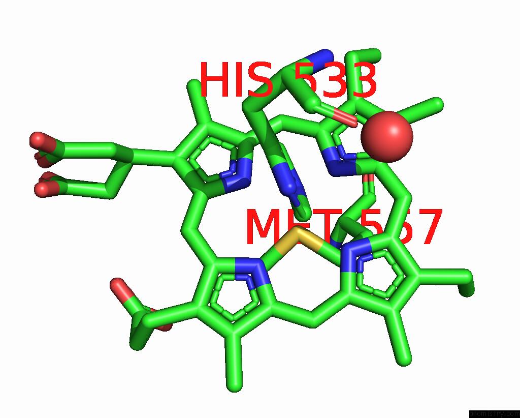
Mono view
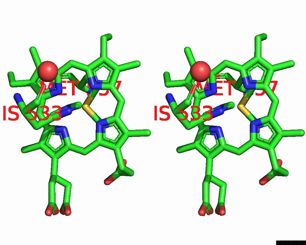
Stereo pair view
Mono view
Stereo pair view
A full contact list of Iron with other atoms in the Fe binding
site number 1 of Sulfite Dehydrogenase From Starkeya Novella Mutant within 5.0Å range:
|
Reference:
S.Bailey,
T.Rapson,
K.Johnson-Winters,
A.V.Astashkin,
J.H.Enemark,
U.Kappler.
Molecular Basis For Enzymatic Sulfite Oxidation: How Three Conserved Active Site Residues Shape Enzyme Activity. J.Biol.Chem. V. 284 2053 2009.
ISSN: ISSN 0021-9258
PubMed: 19004819
DOI: 10.1074/JBC.M807718200
Page generated: Sat Aug 3 20:08:28 2024
ISSN: ISSN 0021-9258
PubMed: 19004819
DOI: 10.1074/JBC.M807718200
Last articles
Zn in 9MJ5Zn in 9HNW
Zn in 9G0L
Zn in 9FNE
Zn in 9DZN
Zn in 9E0I
Zn in 9D32
Zn in 9DAK
Zn in 8ZXC
Zn in 8ZUF