Iron »
PDB 2c2c-2ciy »
2cag »
Iron in PDB 2cag: Catalase Compound II
Enzymatic activity of Catalase Compound II
All present enzymatic activity of Catalase Compound II:
1.11.1.6;
1.11.1.6;
Protein crystallography data
The structure of Catalase Compound II, PDB code: 2cag
was solved by
P.Gouet,
H.M.Jouve,
J.Hajdu,
with X-Ray Crystallography technique. A brief refinement statistics is given in the table below:
| Resolution Low / High (Å) | 15.00 / 2.70 |
| Space group | P 62 2 2 |
| Cell size a, b, c (Å), α, β, γ (°) | 111.840, 111.840, 249.910, 90.00, 90.00, 120.00 |
| R / Rfree (%) | 17.9 / 24.1 |
Iron Binding Sites:
The binding sites of Iron atom in the Catalase Compound II
(pdb code 2cag). This binding sites where shown within
5.0 Angstroms radius around Iron atom.
In total only one binding site of Iron was determined in the Catalase Compound II, PDB code: 2cag:
In total only one binding site of Iron was determined in the Catalase Compound II, PDB code: 2cag:
Iron binding site 1 out of 1 in 2cag
Go back to
Iron binding site 1 out
of 1 in the Catalase Compound II
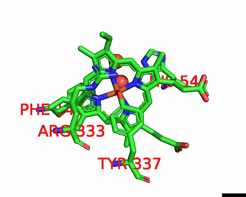
Mono view
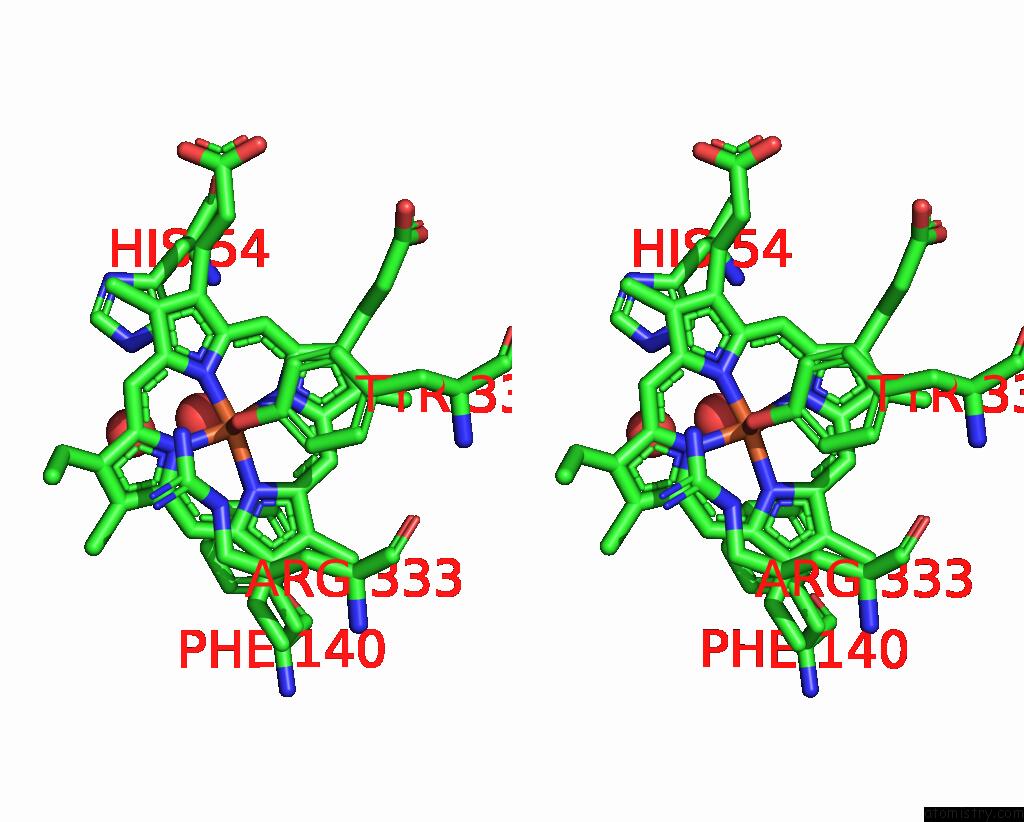
Stereo pair view
Mono view
Stereo pair view
A full contact list of Iron with other atoms in the Fe binding
site number 1 of Catalase Compound II within 5.0Å range:
|
Reference:
P.Gouet,
H.M.Jouve,
P.A.Williams,
I.Andersson,
P.Andreoletti,
L.Nussaume,
J.Hajdu.
Ferryl Intermediates of Catalase Captured By Time-Resolved Weissenberg Crystallography and Uv-Vis Spectroscopy. Nat.Struct.Biol. V. 3 951 1996.
ISSN: ISSN 1072-8368
PubMed: 8901874
DOI: 10.1038/NSB1196-951
Page generated: Thu Jul 17 00:21:37 2025
ISSN: ISSN 1072-8368
PubMed: 8901874
DOI: 10.1038/NSB1196-951
Last articles
Fe in 2YXOFe in 2YRS
Fe in 2YXC
Fe in 2YNM
Fe in 2YVJ
Fe in 2YP1
Fe in 2YU2
Fe in 2YU1
Fe in 2YQB
Fe in 2YOO