Iron »
PDB 2c2c-2ciy »
2cah »
Iron in PDB 2cah: Structure of Proteus Mirabilis Pr Catalase For the Native Form (E- Fe(III)) Complexed with Nadph
Enzymatic activity of Structure of Proteus Mirabilis Pr Catalase For the Native Form (E- Fe(III)) Complexed with Nadph
All present enzymatic activity of Structure of Proteus Mirabilis Pr Catalase For the Native Form (E- Fe(III)) Complexed with Nadph:
1.11.1.6;
1.11.1.6;
Protein crystallography data
The structure of Structure of Proteus Mirabilis Pr Catalase For the Native Form (E- Fe(III)) Complexed with Nadph, PDB code: 2cah
was solved by
P.Gouet,
H.-M.Jouve,
O.Dideberg,
with X-Ray Crystallography technique. A brief refinement statistics is given in the table below:
| Resolution Low / High (Å) | 15.00 / 2.70 |
| Space group | P 62 2 2 |
| Cell size a, b, c (Å), α, β, γ (°) | 112.360, 112.360, 249.140, 90.00, 90.00, 120.00 |
| R / Rfree (%) | 19.6 / n/a |
Iron Binding Sites:
The binding sites of Iron atom in the Structure of Proteus Mirabilis Pr Catalase For the Native Form (E- Fe(III)) Complexed with Nadph
(pdb code 2cah). This binding sites where shown within
5.0 Angstroms radius around Iron atom.
In total only one binding site of Iron was determined in the Structure of Proteus Mirabilis Pr Catalase For the Native Form (E- Fe(III)) Complexed with Nadph, PDB code: 2cah:
In total only one binding site of Iron was determined in the Structure of Proteus Mirabilis Pr Catalase For the Native Form (E- Fe(III)) Complexed with Nadph, PDB code: 2cah:
Iron binding site 1 out of 1 in 2cah
Go back to
Iron binding site 1 out
of 1 in the Structure of Proteus Mirabilis Pr Catalase For the Native Form (E- Fe(III)) Complexed with Nadph
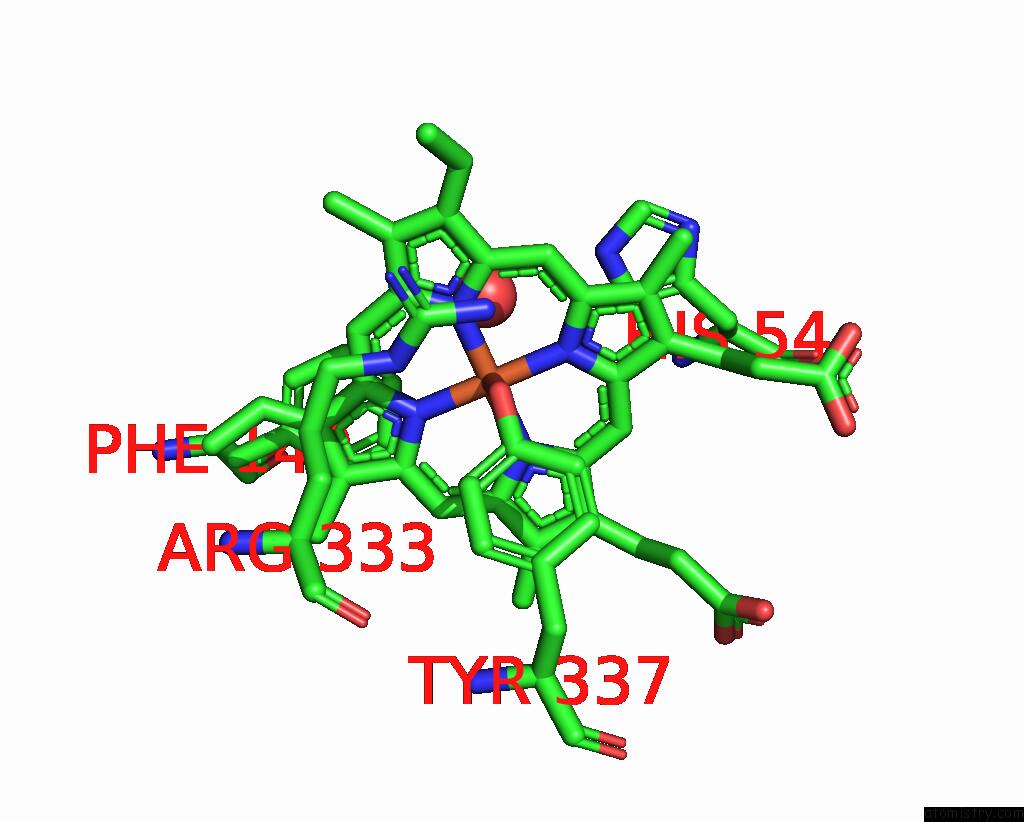
Mono view
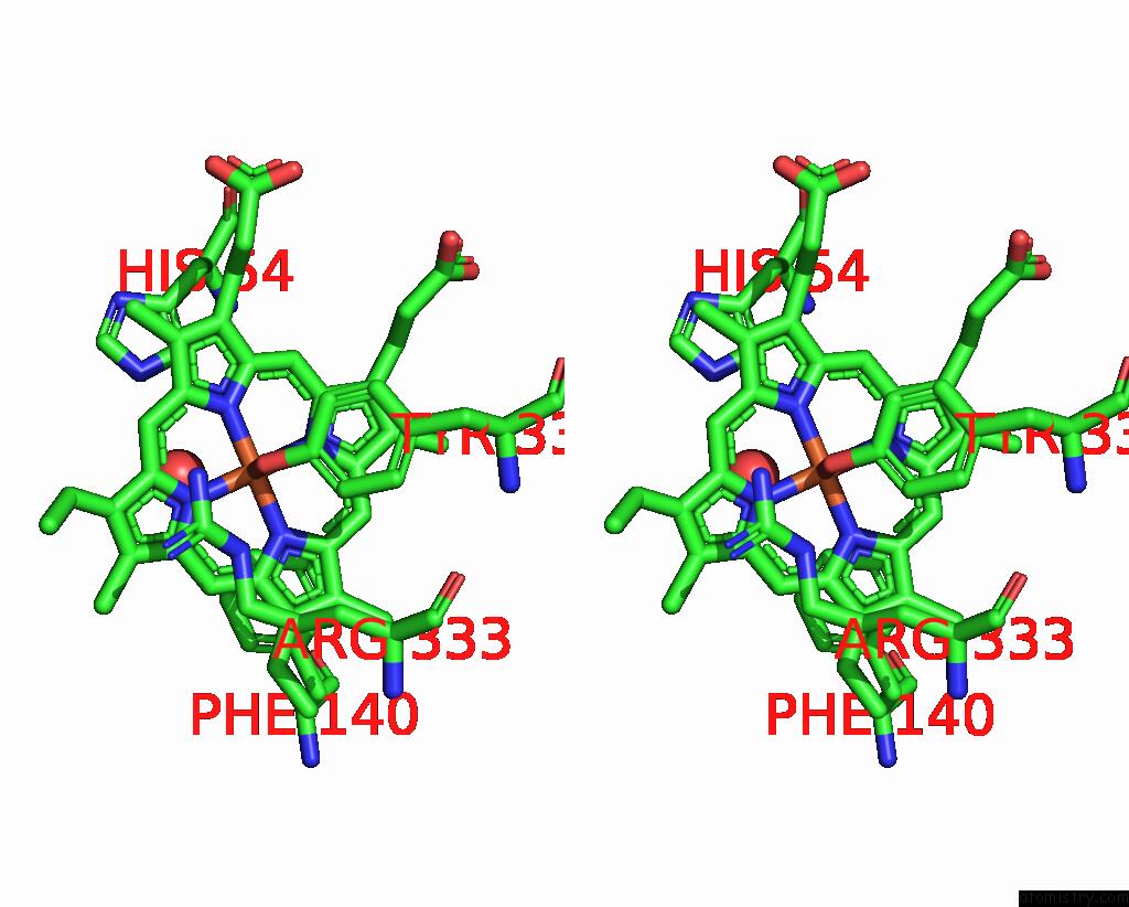
Stereo pair view
Mono view
Stereo pair view
A full contact list of Iron with other atoms in the Fe binding
site number 1 of Structure of Proteus Mirabilis Pr Catalase For the Native Form (E- Fe(III)) Complexed with Nadph within 5.0Å range:
|
Reference:
P.Gouet,
H.M.Jouve,
O.Dideberg.
Crystal Structure of Proteus Mirabilis Pr Catalase with and Without Bound Nadph. J.Mol.Biol. V. 249 933 1995.
ISSN: ISSN 0022-2836
PubMed: 7791219
DOI: 10.1006/JMBI.1995.0350
Page generated: Thu Jul 17 00:21:54 2025
ISSN: ISSN 0022-2836
PubMed: 7791219
DOI: 10.1006/JMBI.1995.0350
Last articles
Fe in 2YXOFe in 2YRS
Fe in 2YXC
Fe in 2YNM
Fe in 2YVJ
Fe in 2YP1
Fe in 2YU2
Fe in 2YU1
Fe in 2YQB
Fe in 2YOO