Iron »
PDB 2c2c-2ciy »
2ciy »
Iron in PDB 2ciy: Chloroperoxidase Complexed with Cyanide and Dmso
Enzymatic activity of Chloroperoxidase Complexed with Cyanide and Dmso
All present enzymatic activity of Chloroperoxidase Complexed with Cyanide and Dmso:
1.11.1.10;
1.11.1.10;
Protein crystallography data
The structure of Chloroperoxidase Complexed with Cyanide and Dmso, PDB code: 2ciy
was solved by
K.Kuhnel,
W.Blankenfeldt,
J.Terner,
I.Schlichting,
with X-Ray Crystallography technique. A brief refinement statistics is given in the table below:
| Resolution Low / High (Å) | 19.84 / 1.70 |
| Space group | C 2 2 21 |
| Cell size a, b, c (Å), α, β, γ (°) | 58.210, 151.030, 101.110, 90.00, 90.00, 90.00 |
| R / Rfree (%) | 17.9 / 20.4 |
Other elements in 2ciy:
The structure of Chloroperoxidase Complexed with Cyanide and Dmso also contains other interesting chemical elements:
| Bromine | (Br) | 2 atoms |
| Manganese | (Mn) | 1 atom |
Iron Binding Sites:
The binding sites of Iron atom in the Chloroperoxidase Complexed with Cyanide and Dmso
(pdb code 2ciy). This binding sites where shown within
5.0 Angstroms radius around Iron atom.
In total only one binding site of Iron was determined in the Chloroperoxidase Complexed with Cyanide and Dmso, PDB code: 2ciy:
In total only one binding site of Iron was determined in the Chloroperoxidase Complexed with Cyanide and Dmso, PDB code: 2ciy:
Iron binding site 1 out of 1 in 2ciy
Go back to
Iron binding site 1 out
of 1 in the Chloroperoxidase Complexed with Cyanide and Dmso
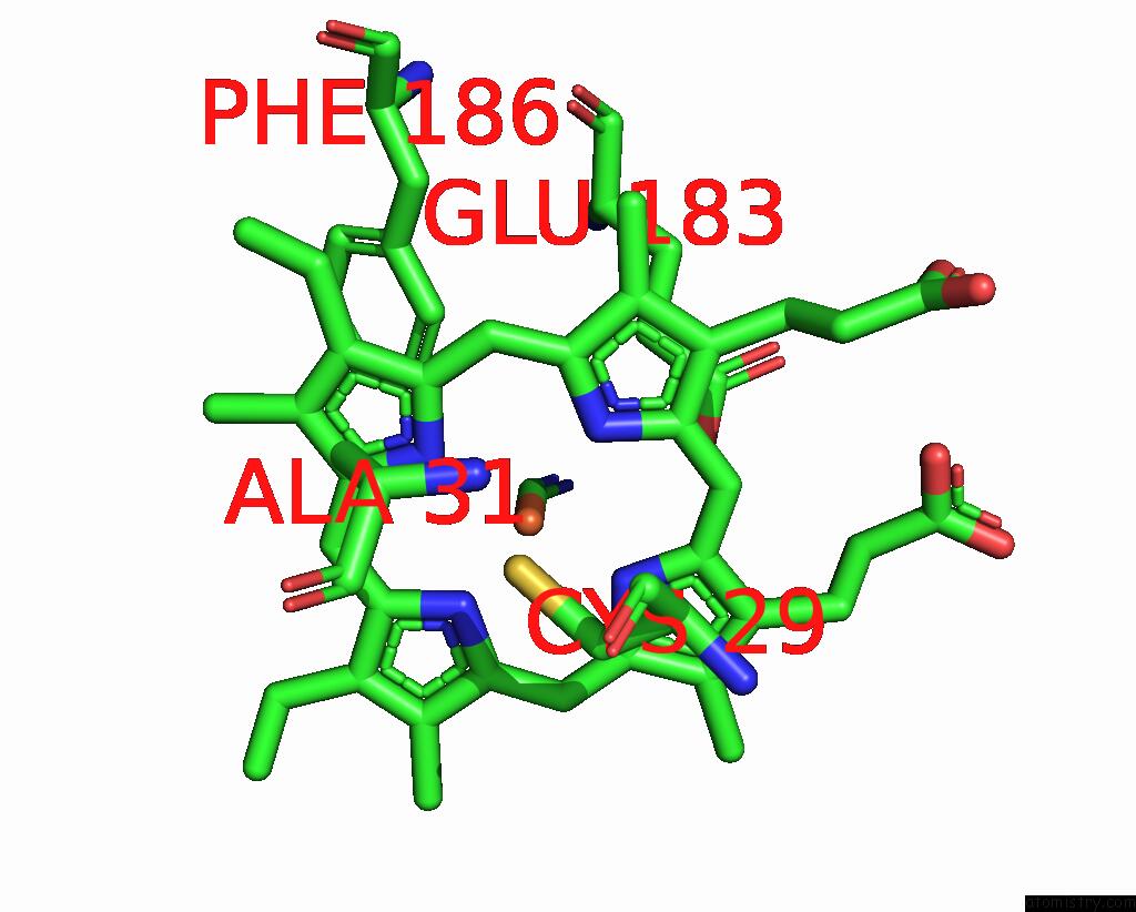
Mono view
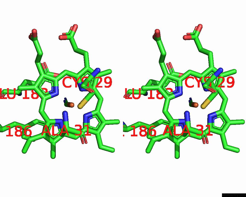
Stereo pair view
Mono view
Stereo pair view
A full contact list of Iron with other atoms in the Fe binding
site number 1 of Chloroperoxidase Complexed with Cyanide and Dmso within 5.0Å range:
|
Reference:
K.Kuhnel,
W.Blankenfeldt,
J.Terner,
I.Schlichting.
Crystal Structures of Chloroperoxidase with Its Bound Substrates and Complexed with Formate, Acetate, and Nitrate. J.Biol.Chem. V. 281 23990 2006.
ISSN: ISSN 0021-9258
PubMed: 16790441
DOI: 10.1074/JBC.M603166200
Page generated: Thu Jul 17 00:26:47 2025
ISSN: ISSN 0021-9258
PubMed: 16790441
DOI: 10.1074/JBC.M603166200
Last articles
Fe in 2YXOFe in 2YRS
Fe in 2YXC
Fe in 2YNM
Fe in 2YVJ
Fe in 2YP1
Fe in 2YU2
Fe in 2YU1
Fe in 2YQB
Fe in 2YOO