Iron »
PDB 2ciz-2d3q »
2cyp »
Iron in PDB 2cyp: Crystal Structure of Yeast Cytochrome C Peroxidase Refined at 1.7- Angstroms Resolution
Enzymatic activity of Crystal Structure of Yeast Cytochrome C Peroxidase Refined at 1.7- Angstroms Resolution
All present enzymatic activity of Crystal Structure of Yeast Cytochrome C Peroxidase Refined at 1.7- Angstroms Resolution:
1.11.1.5;
1.11.1.5;
Protein crystallography data
The structure of Crystal Structure of Yeast Cytochrome C Peroxidase Refined at 1.7- Angstroms Resolution, PDB code: 2cyp
was solved by
B.C.Finzel,
T.L.Poulos,
J.Kraut,
with X-Ray Crystallography technique. A brief refinement statistics is given in the table below:
| Resolution Low / High (Å) | N/A / 1.70 |
| Space group | P 21 21 21 |
| Cell size a, b, c (Å), α, β, γ (°) | 107.400, 76.800, 51.400, 90.00, 90.00, 90.00 |
| R / Rfree (%) | n/a / n/a |
Iron Binding Sites:
The binding sites of Iron atom in the Crystal Structure of Yeast Cytochrome C Peroxidase Refined at 1.7- Angstroms Resolution
(pdb code 2cyp). This binding sites where shown within
5.0 Angstroms radius around Iron atom.
In total only one binding site of Iron was determined in the Crystal Structure of Yeast Cytochrome C Peroxidase Refined at 1.7- Angstroms Resolution, PDB code: 2cyp:
In total only one binding site of Iron was determined in the Crystal Structure of Yeast Cytochrome C Peroxidase Refined at 1.7- Angstroms Resolution, PDB code: 2cyp:
Iron binding site 1 out of 1 in 2cyp
Go back to
Iron binding site 1 out
of 1 in the Crystal Structure of Yeast Cytochrome C Peroxidase Refined at 1.7- Angstroms Resolution
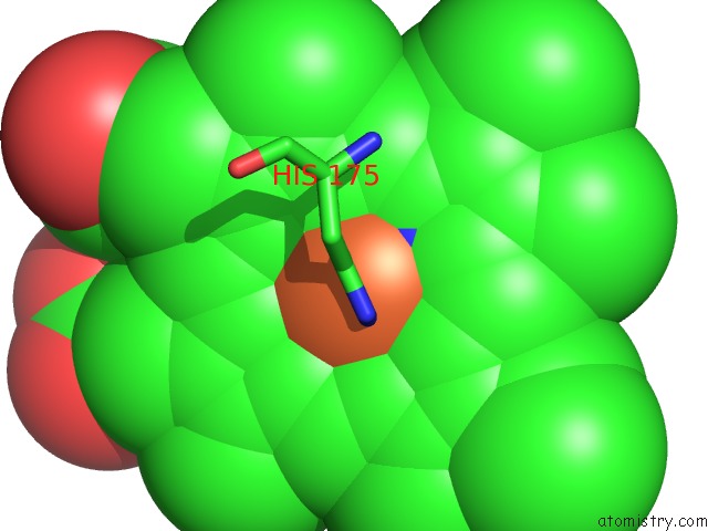
Mono view
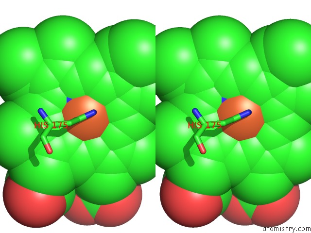
Stereo pair view
Mono view
Stereo pair view
A full contact list of Iron with other atoms in the Fe binding
site number 1 of Crystal Structure of Yeast Cytochrome C Peroxidase Refined at 1.7- Angstroms Resolution within 5.0Å range:
|
Reference:
B.C.Finzel,
T.L.Poulos,
J.Kraut.
Crystal Structure of Yeast Cytochrome C Peroxidase Refined at 1.7-A Resolution J.Biol.Chem. V. 259 13027 1984.
ISSN: ISSN 0021-9258
PubMed: 6092361
Page generated: Sat Aug 3 20:33:19 2024
ISSN: ISSN 0021-9258
PubMed: 6092361
Last articles
Zn in 9J0NZn in 9J0O
Zn in 9J0P
Zn in 9FJX
Zn in 9EKB
Zn in 9C0F
Zn in 9CAH
Zn in 9CH0
Zn in 9CH3
Zn in 9CH1