Iron »
PDB 2d3y-2e1q »
2d3y »
Iron in PDB 2d3y: Crystal Structure of Uracil-Dna Glycosylase From Thermus Thermophilus HB8
Protein crystallography data
The structure of Crystal Structure of Uracil-Dna Glycosylase From Thermus Thermophilus HB8, PDB code: 2d3y
was solved by
H.Kosaka,
N.Nakagawa,
R.Masui,
S.Kuramitsu,
Riken Structuralgenomics/Proteomics Initiative (Rsgi),
with X-Ray Crystallography technique. A brief refinement statistics is given in the table below:
| Resolution Low / High (Å) | 50.00 / 1.55 |
| Space group | P 21 21 21 |
| Cell size a, b, c (Å), α, β, γ (°) | 48.180, 62.350, 63.040, 90.00, 90.00, 90.00 |
| R / Rfree (%) | 18.6 / 21 |
Iron Binding Sites:
The binding sites of Iron atom in the Crystal Structure of Uracil-Dna Glycosylase From Thermus Thermophilus HB8
(pdb code 2d3y). This binding sites where shown within
5.0 Angstroms radius around Iron atom.
In total 4 binding sites of Iron where determined in the Crystal Structure of Uracil-Dna Glycosylase From Thermus Thermophilus HB8, PDB code: 2d3y:
Jump to Iron binding site number: 1; 2; 3; 4;
In total 4 binding sites of Iron where determined in the Crystal Structure of Uracil-Dna Glycosylase From Thermus Thermophilus HB8, PDB code: 2d3y:
Jump to Iron binding site number: 1; 2; 3; 4;
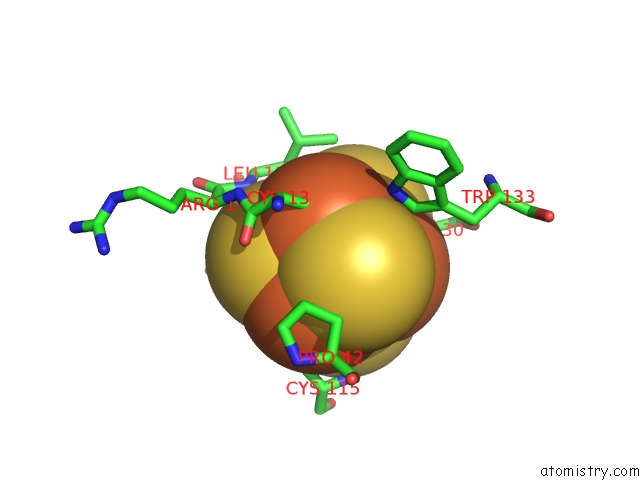
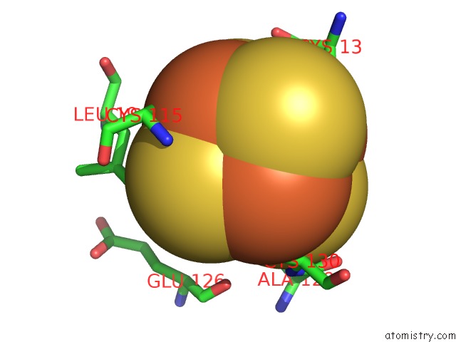
Iron binding site 1 out of 4 in 2d3y
Go back to
Iron binding site 1 out
of 4 in the Crystal Structure of Uracil-Dna Glycosylase From Thermus Thermophilus HB8
Mono view
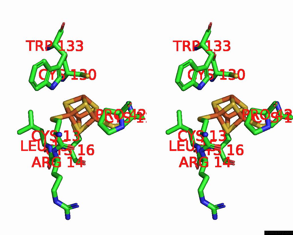
Stereo pair view
Mono view
Stereo pair view
A full contact list of Iron with other atoms in the Fe binding
site number 1 of Crystal Structure of Uracil-Dna Glycosylase From Thermus Thermophilus HB8 within 5.0Å range:
|
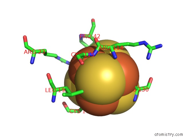
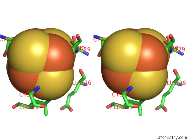
Iron binding site 2 out of 4 in 2d3y
Go back to
Iron binding site 2 out
of 4 in the Crystal Structure of Uracil-Dna Glycosylase From Thermus Thermophilus HB8
Mono view
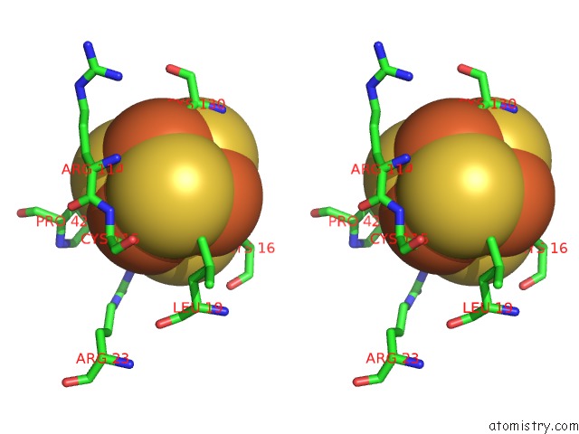
Stereo pair view
Mono view
Stereo pair view
A full contact list of Iron with other atoms in the Fe binding
site number 2 of Crystal Structure of Uracil-Dna Glycosylase From Thermus Thermophilus HB8 within 5.0Å range:
|
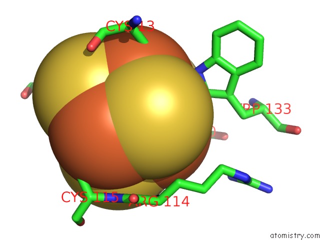
Iron binding site 3 out of 4 in 2d3y
Go back to
Iron binding site 3 out
of 4 in the Crystal Structure of Uracil-Dna Glycosylase From Thermus Thermophilus HB8
Mono view
Stereo pair view
Mono view
Stereo pair view
A full contact list of Iron with other atoms in the Fe binding
site number 3 of Crystal Structure of Uracil-Dna Glycosylase From Thermus Thermophilus HB8 within 5.0Å range:
|
Iron binding site 4 out of 4 in 2d3y
Go back to
Iron binding site 4 out
of 4 in the Crystal Structure of Uracil-Dna Glycosylase From Thermus Thermophilus HB8
Mono view
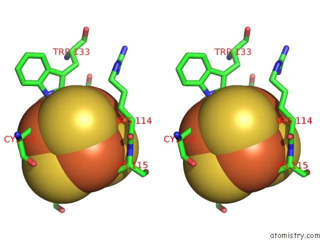
Stereo pair view
Mono view
Stereo pair view
A full contact list of Iron with other atoms in the Fe binding
site number 4 of Crystal Structure of Uracil-Dna Glycosylase From Thermus Thermophilus HB8 within 5.0Å range:
|
Reference:
H.Kosaka,
J.Hoseki,
N.Nakagawa,
S.Kuramitsu,
R.Masui.
Crystal Structure of Family 5 Uracil-Dna Glycosylase Bound to Dna. J.Mol.Biol. V. 373 839 2007.
ISSN: ISSN 0022-2836
PubMed: 17870091
DOI: 10.1016/J.JMB.2007.08.022
Page generated: Sat Aug 3 20:43:20 2024
ISSN: ISSN 0022-2836
PubMed: 17870091
DOI: 10.1016/J.JMB.2007.08.022
Last articles
F in 7LZWF in 7LY8
F in 7LWG
F in 7LZV
F in 7LZF
F in 7LZD
F in 7LZA
F in 7LVX
F in 7LUN
F in 7LVR