Iron »
PDB 2d3y-2e1q »
2dp6 »
Iron in PDB 2dp6: Crystal Structure of Uracil-Dna Glycosylase in Complex with Ap:C Containing Dna
Protein crystallography data
The structure of Crystal Structure of Uracil-Dna Glycosylase in Complex with Ap:C Containing Dna, PDB code: 2dp6
was solved by
H.Kosaka,
N.Nakagawa,
R.Masui,
S.Kuramitsu,
J.Hoseki,
Riken Structuralgenomics/Proteomics Initiative (Rsgi),
with X-Ray Crystallography technique. A brief refinement statistics is given in the table below:
| Resolution Low / High (Å) | 31.25 / 1.80 |
| Space group | C 2 2 21 |
| Cell size a, b, c (Å), α, β, γ (°) | 68.868, 148.754, 93.086, 90.00, 90.00, 90.00 |
| R / Rfree (%) | 20.4 / 21.7 |
Iron Binding Sites:
The binding sites of Iron atom in the Crystal Structure of Uracil-Dna Glycosylase in Complex with Ap:C Containing Dna
(pdb code 2dp6). This binding sites where shown within
5.0 Angstroms radius around Iron atom.
In total 4 binding sites of Iron where determined in the Crystal Structure of Uracil-Dna Glycosylase in Complex with Ap:C Containing Dna, PDB code: 2dp6:
Jump to Iron binding site number: 1; 2; 3; 4;
In total 4 binding sites of Iron where determined in the Crystal Structure of Uracil-Dna Glycosylase in Complex with Ap:C Containing Dna, PDB code: 2dp6:
Jump to Iron binding site number: 1; 2; 3; 4;
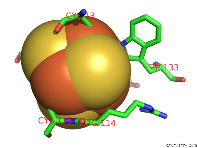
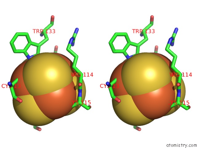
Iron binding site 1 out of 4 in 2dp6
Go back to
Iron binding site 1 out
of 4 in the Crystal Structure of Uracil-Dna Glycosylase in Complex with Ap:C Containing Dna
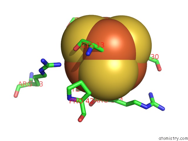
Mono view
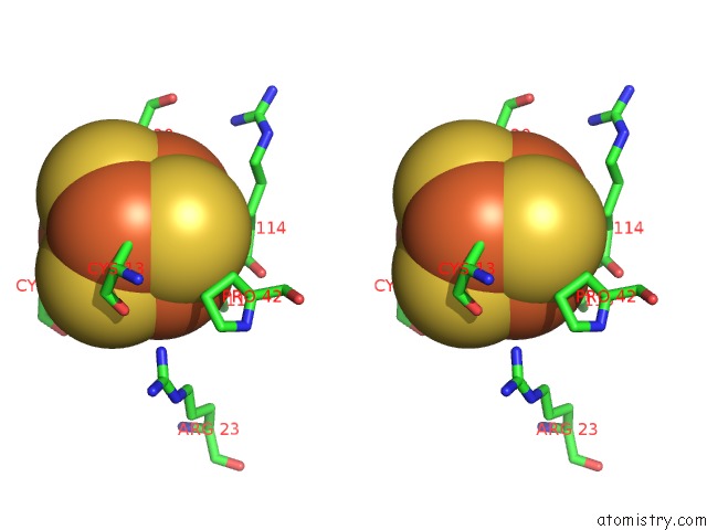
Stereo pair view
Mono view
Stereo pair view
A full contact list of Iron with other atoms in the Fe binding
site number 1 of Crystal Structure of Uracil-Dna Glycosylase in Complex with Ap:C Containing Dna within 5.0Å range:
|
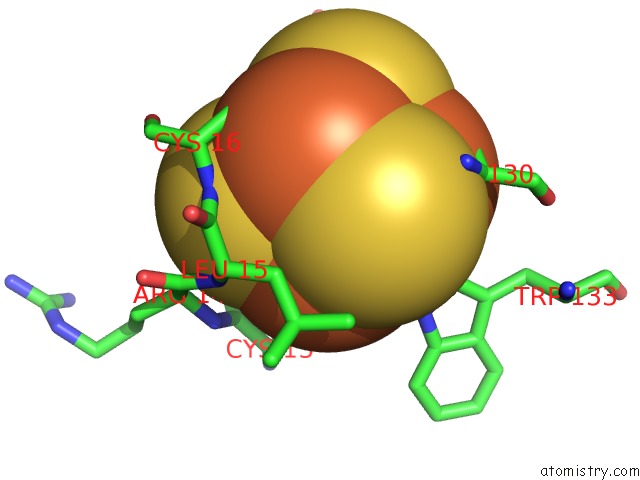
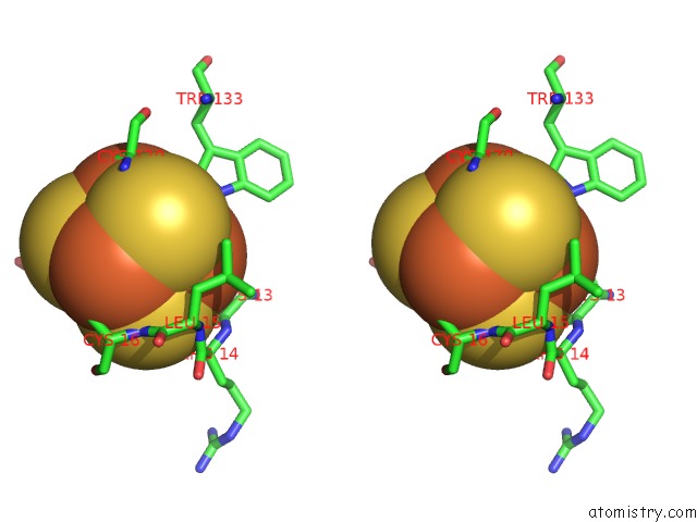
Iron binding site 2 out of 4 in 2dp6
Go back to
Iron binding site 2 out
of 4 in the Crystal Structure of Uracil-Dna Glycosylase in Complex with Ap:C Containing Dna
Mono view
Stereo pair view
Mono view
Stereo pair view
A full contact list of Iron with other atoms in the Fe binding
site number 2 of Crystal Structure of Uracil-Dna Glycosylase in Complex with Ap:C Containing Dna within 5.0Å range:
|
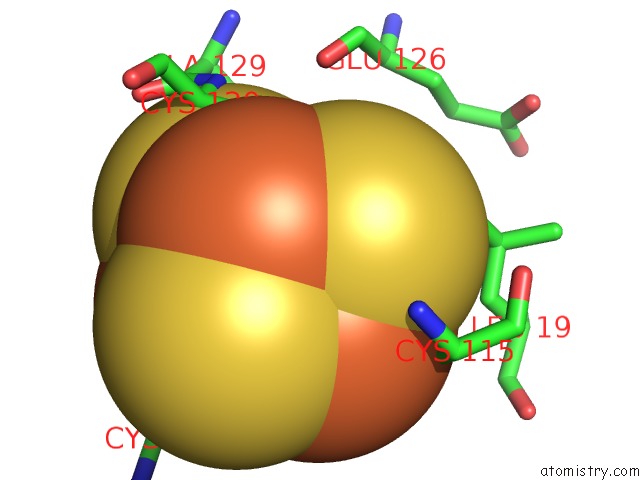
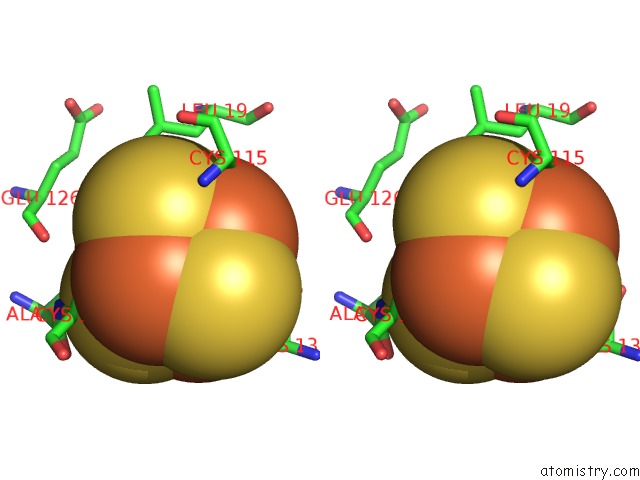
Iron binding site 3 out of 4 in 2dp6
Go back to
Iron binding site 3 out
of 4 in the Crystal Structure of Uracil-Dna Glycosylase in Complex with Ap:C Containing Dna
Mono view
Stereo pair view
Mono view
Stereo pair view
A full contact list of Iron with other atoms in the Fe binding
site number 3 of Crystal Structure of Uracil-Dna Glycosylase in Complex with Ap:C Containing Dna within 5.0Å range:
|
Iron binding site 4 out of 4 in 2dp6
Go back to
Iron binding site 4 out
of 4 in the Crystal Structure of Uracil-Dna Glycosylase in Complex with Ap:C Containing Dna
Mono view
Stereo pair view
Mono view
Stereo pair view
A full contact list of Iron with other atoms in the Fe binding
site number 4 of Crystal Structure of Uracil-Dna Glycosylase in Complex with Ap:C Containing Dna within 5.0Å range:
|
Reference:
H.Kosaka,
N.Nakagawa,
R.Masui,
S.Kuramitsu.
Structure of Family 5 Uracil-Dna Glycosylase Bound to Dna Reveals Insights Into the Mechanism For Substrate Recognition and Catalysis To Be Published.
Page generated: Thu Jul 17 00:44:09 2025
Last articles
Fe in 2YXOFe in 2YRS
Fe in 2YXC
Fe in 2YNM
Fe in 2YVJ
Fe in 2YP1
Fe in 2YU2
Fe in 2YU1
Fe in 2YQB
Fe in 2YOO