Iron »
PDB 2d3y-2e1q »
2dxr »
Iron in PDB 2dxr: Crystal Structure of the Complex Formed Between C-Terminal Half of Bovine Lactoferrin and Sorbitol at 2.85 A Resolution
Protein crystallography data
The structure of Crystal Structure of the Complex Formed Between C-Terminal Half of Bovine Lactoferrin and Sorbitol at 2.85 A Resolution, PDB code: 2dxr
was solved by
R.Mir,
R.Prem Kumar,
M.Sinha,
N.Singh,
S.Sharma,
A.Bhushan,
P.Kaur,
T.P.Singh,
with X-Ray Crystallography technique. A brief refinement statistics is given in the table below:
| Resolution Low / High (Å) | 20.00 / 2.85 |
| Space group | P 1 21 1 |
| Cell size a, b, c (Å), α, β, γ (°) | 63.382, 50.391, 65.856, 90.00, 107.78, 90.00 |
| R / Rfree (%) | 19.8 / 22.7 |
Other elements in 2dxr:
The structure of Crystal Structure of the Complex Formed Between C-Terminal Half of Bovine Lactoferrin and Sorbitol at 2.85 A Resolution also contains other interesting chemical elements:
| Zinc | (Zn) | 2 atoms |
Iron Binding Sites:
The binding sites of Iron atom in the Crystal Structure of the Complex Formed Between C-Terminal Half of Bovine Lactoferrin and Sorbitol at 2.85 A Resolution
(pdb code 2dxr). This binding sites where shown within
5.0 Angstroms radius around Iron atom.
In total only one binding site of Iron was determined in the Crystal Structure of the Complex Formed Between C-Terminal Half of Bovine Lactoferrin and Sorbitol at 2.85 A Resolution, PDB code: 2dxr:
In total only one binding site of Iron was determined in the Crystal Structure of the Complex Formed Between C-Terminal Half of Bovine Lactoferrin and Sorbitol at 2.85 A Resolution, PDB code: 2dxr:
Iron binding site 1 out of 1 in 2dxr
Go back to
Iron binding site 1 out
of 1 in the Crystal Structure of the Complex Formed Between C-Terminal Half of Bovine Lactoferrin and Sorbitol at 2.85 A Resolution
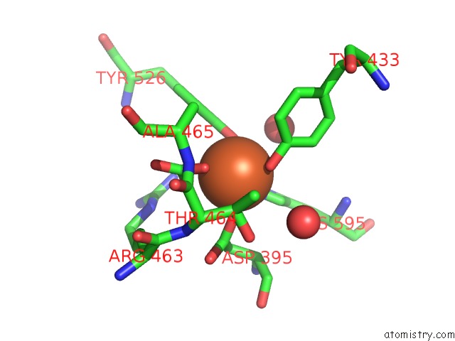
Mono view
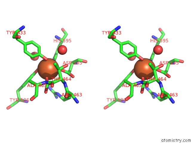
Stereo pair view
Mono view
Stereo pair view
A full contact list of Iron with other atoms in the Fe binding
site number 1 of Crystal Structure of the Complex Formed Between C-Terminal Half of Bovine Lactoferrin and Sorbitol at 2.85 A Resolution within 5.0Å range:
|
Reference:
R.Mir,
R.Prem Kumar,
M.Sinha,
N.Singh,
S.Sharma,
A.Bhushan,
P.Kaur,
T.P.Singh.
Crystal Structure of the Complex Formed Between C-Terminal Half of Bovine Lactoferrin and Sorbitol at 2.85 A Resolution To Be Published.
Page generated: Thu Jul 17 00:45:30 2025
Last articles
Fe in 2YXOFe in 2YRS
Fe in 2YXC
Fe in 2YNM
Fe in 2YVJ
Fe in 2YP1
Fe in 2YU2
Fe in 2YU1
Fe in 2YQB
Fe in 2YOO