Iron »
PDB 2e1s-2eus »
2e4p »
Iron in PDB 2e4p: Crystal Structure of BPHA3 (Oxidized Form)
Protein crystallography data
The structure of Crystal Structure of BPHA3 (Oxidized Form), PDB code: 2e4p
was solved by
M.Senda,
S.Kishigami,
S.Kimura,
T.Ishida,
M.Fukuda,
T.Senda,
with X-Ray Crystallography technique. A brief refinement statistics is given in the table below:
| Resolution Low / High (Å) | 16.65 / 2.00 |
| Space group | P 21 21 2 |
| Cell size a, b, c (Å), α, β, γ (°) | 26.294, 144.113, 61.176, 90.00, 90.00, 90.00 |
| R / Rfree (%) | 20.8 / 25.4 |
Iron Binding Sites:
The binding sites of Iron atom in the Crystal Structure of BPHA3 (Oxidized Form)
(pdb code 2e4p). This binding sites where shown within
5.0 Angstroms radius around Iron atom.
In total 4 binding sites of Iron where determined in the Crystal Structure of BPHA3 (Oxidized Form), PDB code: 2e4p:
Jump to Iron binding site number: 1; 2; 3; 4;
In total 4 binding sites of Iron where determined in the Crystal Structure of BPHA3 (Oxidized Form), PDB code: 2e4p:
Jump to Iron binding site number: 1; 2; 3; 4;
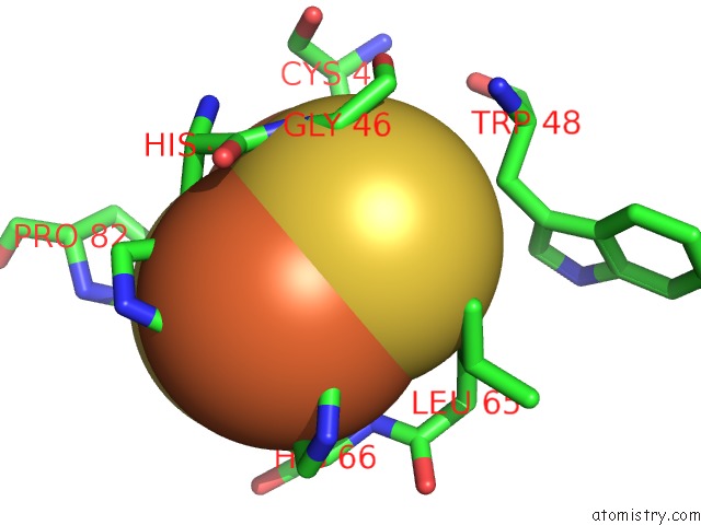
Iron binding site 1 out of 4 in 2e4p
Go back to
Iron binding site 1 out
of 4 in the Crystal Structure of BPHA3 (Oxidized Form)
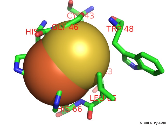
Mono view
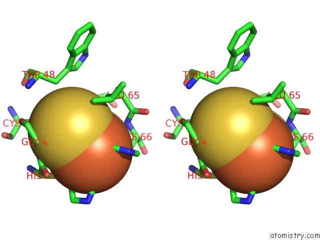
Stereo pair view
Mono view
Stereo pair view
A full contact list of Iron with other atoms in the Fe binding
site number 1 of Crystal Structure of BPHA3 (Oxidized Form) within 5.0Å range:
|
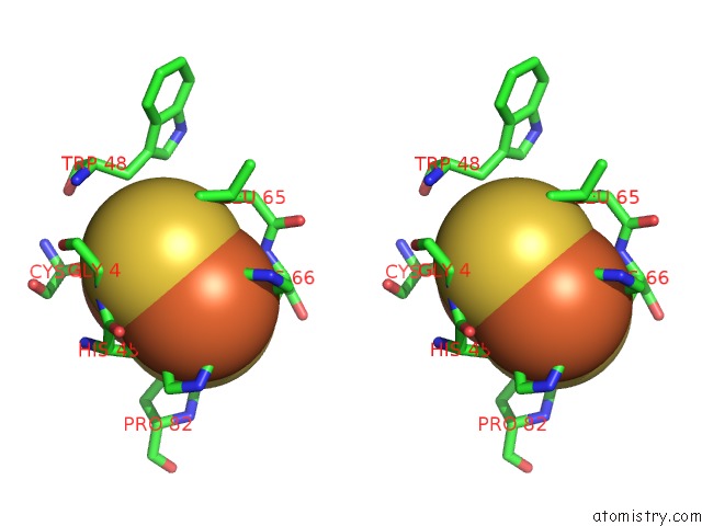
Iron binding site 2 out of 4 in 2e4p
Go back to
Iron binding site 2 out
of 4 in the Crystal Structure of BPHA3 (Oxidized Form)
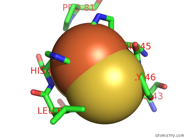
Mono view
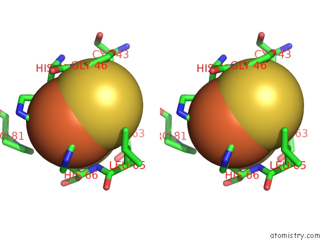
Stereo pair view
Mono view
Stereo pair view
A full contact list of Iron with other atoms in the Fe binding
site number 2 of Crystal Structure of BPHA3 (Oxidized Form) within 5.0Å range:
|
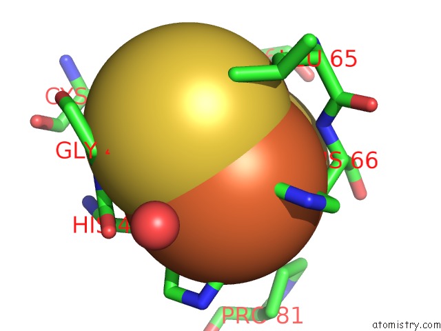
Iron binding site 3 out of 4 in 2e4p
Go back to
Iron binding site 3 out
of 4 in the Crystal Structure of BPHA3 (Oxidized Form)
Mono view
Stereo pair view
Mono view
Stereo pair view
A full contact list of Iron with other atoms in the Fe binding
site number 3 of Crystal Structure of BPHA3 (Oxidized Form) within 5.0Å range:
|
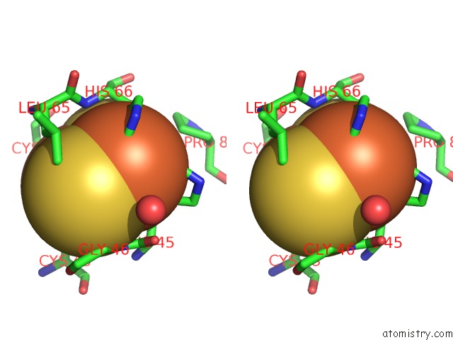
Iron binding site 4 out of 4 in 2e4p
Go back to
Iron binding site 4 out
of 4 in the Crystal Structure of BPHA3 (Oxidized Form)
Mono view
Stereo pair view
Mono view
Stereo pair view
A full contact list of Iron with other atoms in the Fe binding
site number 4 of Crystal Structure of BPHA3 (Oxidized Form) within 5.0Å range:
|
Reference:
M.Senda,
S.Kishigami,
S.Kimura,
M.Fukuda,
T.Ishida,
T.Senda.
Molecular Mechanism of the Redox-Dependent Interaction Between Nadh-Dependent Ferredoxin Reductase and Rieske-Type [2FE-2S] Ferredoxin J.Mol.Biol. V. 373 382 2007.
ISSN: ISSN 0022-2836
PubMed: 17850818
DOI: 10.1016/J.JMB.2007.08.002
Page generated: Thu Jul 17 00:48:22 2025
ISSN: ISSN 0022-2836
PubMed: 17850818
DOI: 10.1016/J.JMB.2007.08.002
Last articles
Fe in 2YXOFe in 2YRS
Fe in 2YXC
Fe in 2YNM
Fe in 2YVJ
Fe in 2YP1
Fe in 2YU2
Fe in 2YU1
Fe in 2YQB
Fe in 2YOO