Iron »
PDB 2e1s-2eus »
2ei2 »
Iron in PDB 2ei2: Crystal Structure Analysis of the 1,2-Dihydroxynaphthalene Dioxygenase From Pseudomonas Sp. Stain C18
Protein crystallography data
The structure of Crystal Structure Analysis of the 1,2-Dihydroxynaphthalene Dioxygenase From Pseudomonas Sp. Stain C18, PDB code: 2ei2
was solved by
D.B.Neau,
M.S.Kelker,
C.L.Colbert,
J.T.Bolin,
with X-Ray Crystallography technique. A brief refinement statistics is given in the table below:
| Resolution Low / High (Å) | 48.97 / 1.69 |
| Space group | I 4 2 2 |
| Cell size a, b, c (Å), α, β, γ (°) | 118.390, 118.390, 120.660, 90.00, 90.00, 90.00 |
| R / Rfree (%) | 16.1 / 18.6 |
Other elements in 2ei2:
The structure of Crystal Structure Analysis of the 1,2-Dihydroxynaphthalene Dioxygenase From Pseudomonas Sp. Stain C18 also contains other interesting chemical elements:
| Magnesium | (Mg) | 1 atom |
Iron Binding Sites:
The binding sites of Iron atom in the Crystal Structure Analysis of the 1,2-Dihydroxynaphthalene Dioxygenase From Pseudomonas Sp. Stain C18
(pdb code 2ei2). This binding sites where shown within
5.0 Angstroms radius around Iron atom.
In total only one binding site of Iron was determined in the Crystal Structure Analysis of the 1,2-Dihydroxynaphthalene Dioxygenase From Pseudomonas Sp. Stain C18, PDB code: 2ei2:
In total only one binding site of Iron was determined in the Crystal Structure Analysis of the 1,2-Dihydroxynaphthalene Dioxygenase From Pseudomonas Sp. Stain C18, PDB code: 2ei2:
Iron binding site 1 out of 1 in 2ei2
Go back to
Iron binding site 1 out
of 1 in the Crystal Structure Analysis of the 1,2-Dihydroxynaphthalene Dioxygenase From Pseudomonas Sp. Stain C18
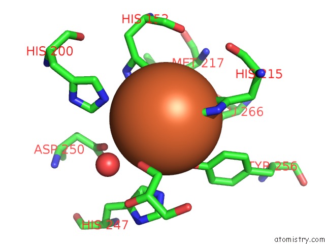
Mono view
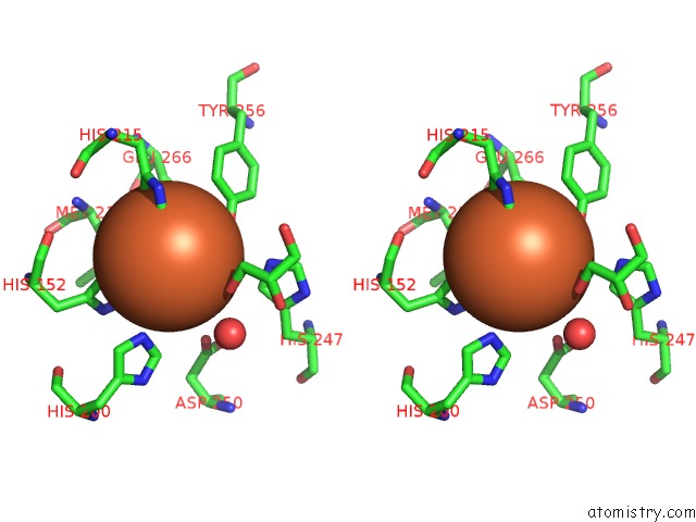
Stereo pair view
Mono view
Stereo pair view
A full contact list of Iron with other atoms in the Fe binding
site number 1 of Crystal Structure Analysis of the 1,2-Dihydroxynaphthalene Dioxygenase From Pseudomonas Sp. Stain C18 within 5.0Å range:
|
Reference:
D.B.Neau,
M.S.Kelker,
H.Maaroufi,
C.L.Colbert,
L.D.Eltis,
J.T.Bolin.
Structural Explanation For Success and Failure in the Enzymatic Ring-Cleavage of 3,4 Dihydroxybiphenyl and Related Pcb Metabolites To Be Published.
Page generated: Thu Jul 17 00:53:41 2025
Last articles
Fe in 2YXOFe in 2YRS
Fe in 2YXC
Fe in 2YNM
Fe in 2YVJ
Fe in 2YP1
Fe in 2YU2
Fe in 2YU1
Fe in 2YQB
Fe in 2YOO