Iron »
PDB 2e1s-2eus »
2ekt »
Iron in PDB 2ekt: Crystal Structure of Myoglobin Reconstituted with 6-Methyl-6- Depropionatehemin
Protein crystallography data
The structure of Crystal Structure of Myoglobin Reconstituted with 6-Methyl-6- Depropionatehemin, PDB code: 2ekt
was solved by
K.Harada,
M.Makino,
H.Sugimoto,
S.Hirota,
T.Matsuo,
Y.Shiro,
Y.Hisaeda,
T.Hayashi,
with X-Ray Crystallography technique. A brief refinement statistics is given in the table below:
| Resolution Low / High (Å) | 10.00 / 1.10 |
| Space group | P 6 |
| Cell size a, b, c (Å), α, β, γ (°) | 90.248, 90.248, 45.346, 90.00, 90.00, 120.00 |
| R / Rfree (%) | 13.1 / 15.8 |
Iron Binding Sites:
The binding sites of Iron atom in the Crystal Structure of Myoglobin Reconstituted with 6-Methyl-6- Depropionatehemin
(pdb code 2ekt). This binding sites where shown within
5.0 Angstroms radius around Iron atom.
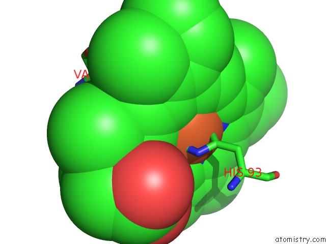
In total only one binding site of Iron was determined in the Crystal Structure of Myoglobin Reconstituted with 6-Methyl-6- Depropionatehemin, PDB code: 2ekt:
In total only one binding site of Iron was determined in the Crystal Structure of Myoglobin Reconstituted with 6-Methyl-6- Depropionatehemin, PDB code: 2ekt:
Iron binding site 1 out of 1 in 2ekt
Go back to
Iron binding site 1 out
of 1 in the Crystal Structure of Myoglobin Reconstituted with 6-Methyl-6- Depropionatehemin
Mono view
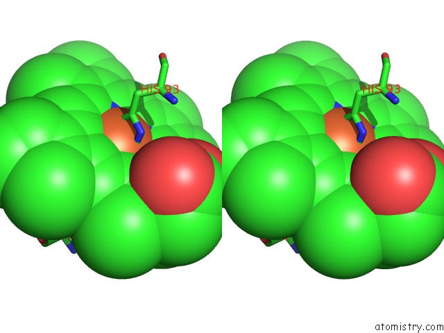
Stereo pair view
Mono view
Stereo pair view
A full contact list of Iron with other atoms in the Fe binding
site number 1 of Crystal Structure of Myoglobin Reconstituted with 6-Methyl-6- Depropionatehemin within 5.0Å range:
|
Reference:
K.Harada,
M.Makino,
H.Sugimoto,
S.Hirota,
T.Matsuo,
Y.Shiro,
Y.Hisaeda,
T.Hayashi.
Structure and Ligand Binding Properties of Myoglobins Reconstituted with Monodepropionated Heme: Functional Role of Each Heme Propionate Side Chain Biochemistry V. 46 9406 2007.
ISSN: ISSN 0006-2960
PubMed: 17636874
DOI: 10.1021/BI7007068
Page generated: Thu Jul 17 00:54:48 2025
ISSN: ISSN 0006-2960
PubMed: 17636874
DOI: 10.1021/BI7007068
Last articles
Fe in 2YXOFe in 2YRS
Fe in 2YXC
Fe in 2YNM
Fe in 2YVJ
Fe in 2YP1
Fe in 2YU2
Fe in 2YU1
Fe in 2YQB
Fe in 2YOO