Iron »
PDB 2e1s-2eus »
2eu7 »
Iron in PDB 2eu7: Crystal Structure of D1A Mutant of Nitrophorin 2 Complexed with Ammonia
Protein crystallography data
The structure of Crystal Structure of D1A Mutant of Nitrophorin 2 Complexed with Ammonia, PDB code: 2eu7
was solved by
A.Weichsel,
W.R.Montfort,
with X-Ray Crystallography technique. A brief refinement statistics is given in the table below:
| Resolution Low / High (Å) | 6.00 / 1.20 |
| Space group | P 21 21 2 |
| Cell size a, b, c (Å), α, β, γ (°) | 39.770, 125.300, 34.080, 90.00, 90.00, 90.00 |
| R / Rfree (%) | 17.7 / 21.5 |
Iron Binding Sites:
The binding sites of Iron atom in the Crystal Structure of D1A Mutant of Nitrophorin 2 Complexed with Ammonia
(pdb code 2eu7). This binding sites where shown within
5.0 Angstroms radius around Iron atom.
In total only one binding site of Iron was determined in the Crystal Structure of D1A Mutant of Nitrophorin 2 Complexed with Ammonia, PDB code: 2eu7:
In total only one binding site of Iron was determined in the Crystal Structure of D1A Mutant of Nitrophorin 2 Complexed with Ammonia, PDB code: 2eu7:
Iron binding site 1 out of 1 in 2eu7
Go back to
Iron binding site 1 out
of 1 in the Crystal Structure of D1A Mutant of Nitrophorin 2 Complexed with Ammonia
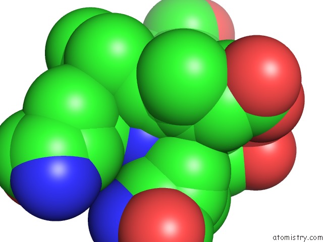
Mono view
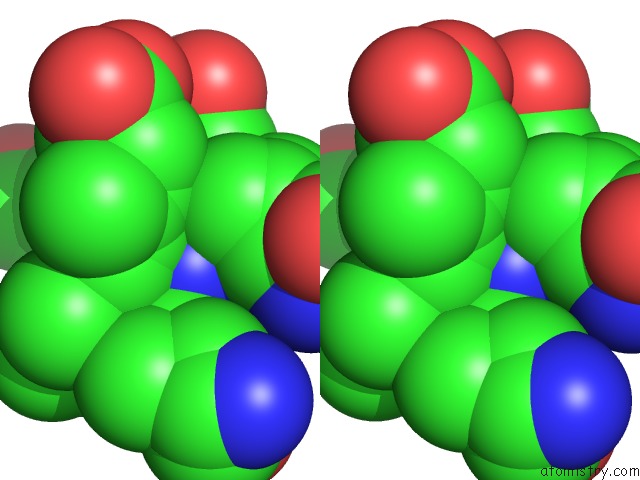
Stereo pair view
Mono view
Stereo pair view
A full contact list of Iron with other atoms in the Fe binding
site number 1 of Crystal Structure of D1A Mutant of Nitrophorin 2 Complexed with Ammonia within 5.0Å range:
|
Reference:
A.Weichsel,
R.E.Berry,
F.A.Walker,
W.R.Montfort.
Crystal Structures, Ligand Induced Conformational Change and Heme Deformation in Complexes of Nitrophorin 2, A Nitric Oxide Transport Protein From Rhodnius Prolixus To Be Published.
Page generated: Thu Jul 17 00:55:20 2025
Last articles
Fe in 2YXOFe in 2YRS
Fe in 2YXC
Fe in 2YNM
Fe in 2YVJ
Fe in 2YP1
Fe in 2YU2
Fe in 2YU1
Fe in 2YQB
Fe in 2YOO