Iron »
PDB 2eut-2fkz »
2fd2 »
Iron in PDB 2fd2: Crystallographic Analysis of Two Site-Directed Mutants of Azotobacter Vinelandii Ferredoxin
Protein crystallography data
The structure of Crystallographic Analysis of Two Site-Directed Mutants of Azotobacter Vinelandii Ferredoxin, PDB code: 2fd2
was solved by
C.D.Stout,
with X-Ray Crystallography technique. A brief refinement statistics is given in the table below:
| Resolution Low / High (Å) | 8.00 / 1.90 |
| Space group | P 41 21 2 |
| Cell size a, b, c (Å), α, β, γ (°) | 55.200, 55.200, 95.200, 90.00, 90.00, 90.00 |
| R / Rfree (%) | 23.2 / n/a |
Iron Binding Sites:
The binding sites of Iron atom in the Crystallographic Analysis of Two Site-Directed Mutants of Azotobacter Vinelandii Ferredoxin
(pdb code 2fd2). This binding sites where shown within
5.0 Angstroms radius around Iron atom.
In total 7 binding sites of Iron where determined in the Crystallographic Analysis of Two Site-Directed Mutants of Azotobacter Vinelandii Ferredoxin, PDB code: 2fd2:
Jump to Iron binding site number: 1; 2; 3; 4; 5; 6; 7;
In total 7 binding sites of Iron where determined in the Crystallographic Analysis of Two Site-Directed Mutants of Azotobacter Vinelandii Ferredoxin, PDB code: 2fd2:
Jump to Iron binding site number: 1; 2; 3; 4; 5; 6; 7;
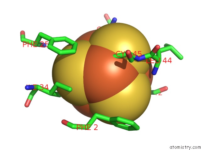
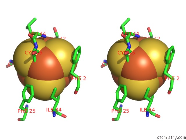
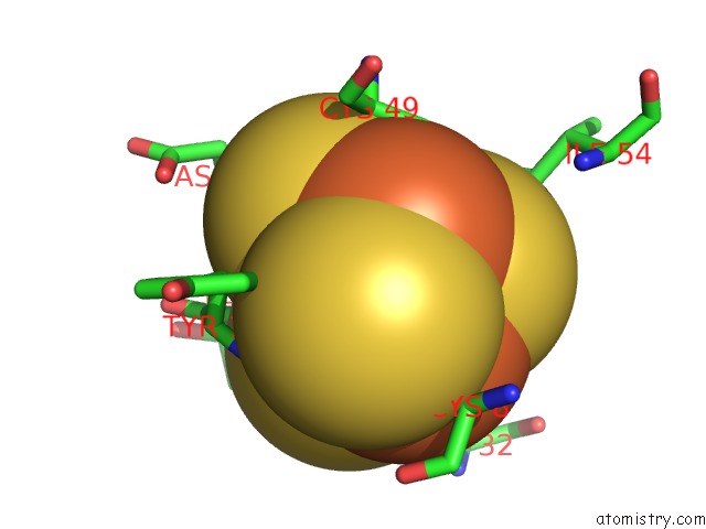
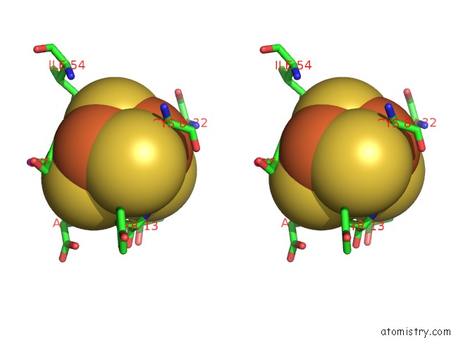
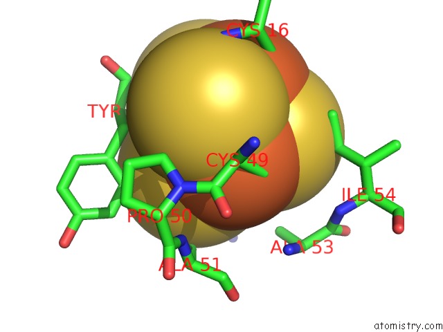
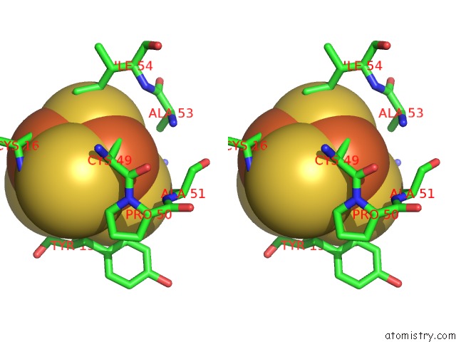
Iron binding site 1 out of 7 in 2fd2
Go back to
Iron binding site 1 out
of 7 in the Crystallographic Analysis of Two Site-Directed Mutants of Azotobacter Vinelandii Ferredoxin
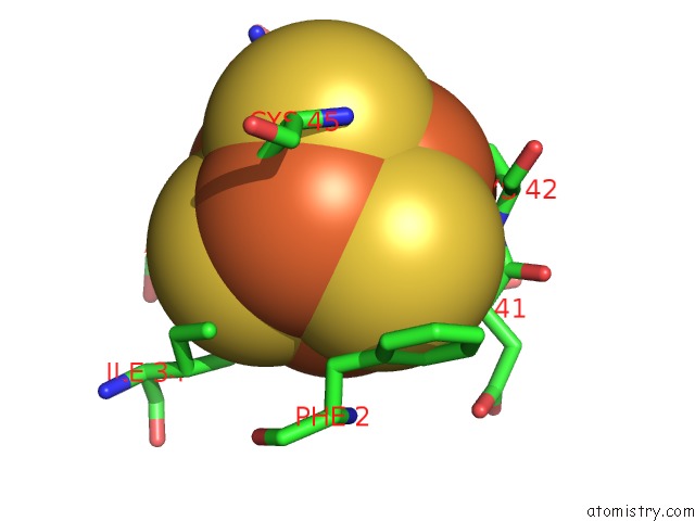
Mono view
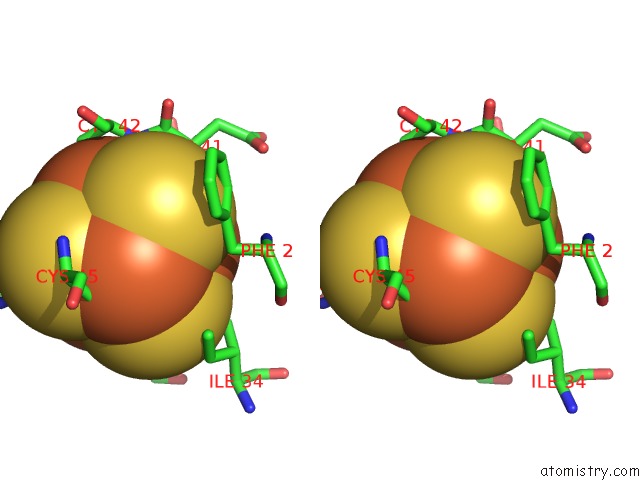
Stereo pair view
Mono view
Stereo pair view
A full contact list of Iron with other atoms in the Fe binding
site number 1 of Crystallographic Analysis of Two Site-Directed Mutants of Azotobacter Vinelandii Ferredoxin within 5.0Å range:
|
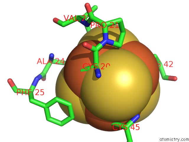
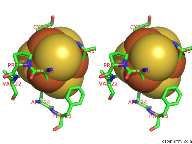
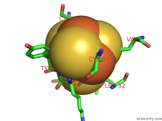
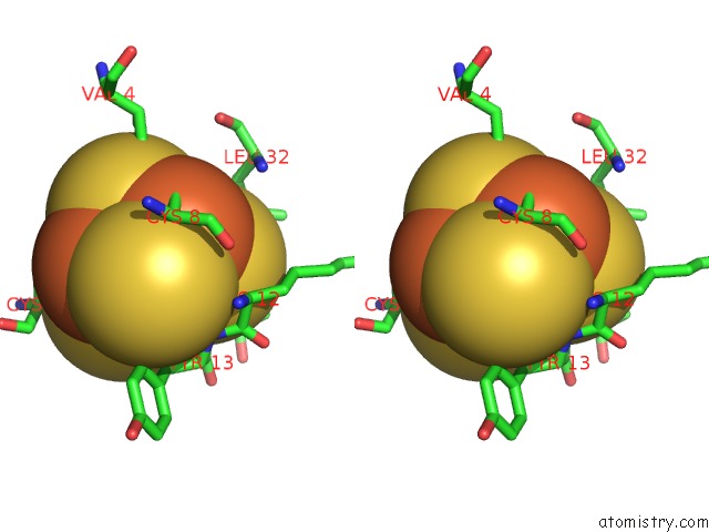
Iron binding site 2 out of 7 in 2fd2
Go back to
Iron binding site 2 out
of 7 in the Crystallographic Analysis of Two Site-Directed Mutants of Azotobacter Vinelandii Ferredoxin
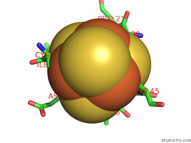
Mono view
Stereo pair view
Mono view
Stereo pair view
A full contact list of Iron with other atoms in the Fe binding
site number 2 of Crystallographic Analysis of Two Site-Directed Mutants of Azotobacter Vinelandii Ferredoxin within 5.0Å range:
|
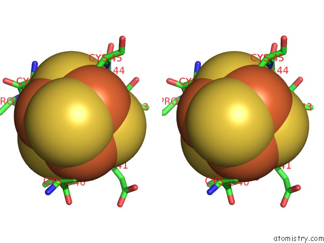
Iron binding site 3 out of 7 in 2fd2
Go back to
Iron binding site 3 out
of 7 in the Crystallographic Analysis of Two Site-Directed Mutants of Azotobacter Vinelandii Ferredoxin
Mono view
Stereo pair view
Mono view
Stereo pair view
A full contact list of Iron with other atoms in the Fe binding
site number 3 of Crystallographic Analysis of Two Site-Directed Mutants of Azotobacter Vinelandii Ferredoxin within 5.0Å range:
|
Iron binding site 4 out of 7 in 2fd2
Go back to
Iron binding site 4 out
of 7 in the Crystallographic Analysis of Two Site-Directed Mutants of Azotobacter Vinelandii Ferredoxin
Mono view
Stereo pair view
Mono view
Stereo pair view
A full contact list of Iron with other atoms in the Fe binding
site number 4 of Crystallographic Analysis of Two Site-Directed Mutants of Azotobacter Vinelandii Ferredoxin within 5.0Å range:
|
Iron binding site 5 out of 7 in 2fd2
Go back to
Iron binding site 5 out
of 7 in the Crystallographic Analysis of Two Site-Directed Mutants of Azotobacter Vinelandii Ferredoxin
Mono view
Stereo pair view
Mono view
Stereo pair view
A full contact list of Iron with other atoms in the Fe binding
site number 5 of Crystallographic Analysis of Two Site-Directed Mutants of Azotobacter Vinelandii Ferredoxin within 5.0Å range:
|
Iron binding site 6 out of 7 in 2fd2
Go back to
Iron binding site 6 out
of 7 in the Crystallographic Analysis of Two Site-Directed Mutants of Azotobacter Vinelandii Ferredoxin
Mono view
Stereo pair view
Mono view
Stereo pair view
A full contact list of Iron with other atoms in the Fe binding
site number 6 of Crystallographic Analysis of Two Site-Directed Mutants of Azotobacter Vinelandii Ferredoxin within 5.0Å range:
|
Iron binding site 7 out of 7 in 2fd2
Go back to
Iron binding site 7 out
of 7 in the Crystallographic Analysis of Two Site-Directed Mutants of Azotobacter Vinelandii Ferredoxin
Mono view
Stereo pair view
Mono view
Stereo pair view
A full contact list of Iron with other atoms in the Fe binding
site number 7 of Crystallographic Analysis of Two Site-Directed Mutants of Azotobacter Vinelandii Ferredoxin within 5.0Å range:
|
Reference:
J.Soman,
S.Iismaa,
C.D.Stout.
Crystallographic Analysis of Two Site-Directed Mutants of Azotobacter Vinelandii Ferredoxin. J.Biol.Chem. V. 266 21558 1991.
ISSN: ISSN 0021-9258
PubMed: 1939185
Page generated: Sat Aug 3 21:16:48 2024
ISSN: ISSN 0021-9258
PubMed: 1939185
Last articles
Zn in 9MJ5Zn in 9HNW
Zn in 9G0L
Zn in 9FNE
Zn in 9DZN
Zn in 9E0I
Zn in 9D32
Zn in 9DAK
Zn in 8ZXC
Zn in 8ZUF