Iron »
PDB 2eut-2fkz »
2fdi »
Iron in PDB 2fdi: Crystal Structure of Alkb in Complex with Fe(II), 2-Oxoglutarate, and Methylated Trinucleotide T-Mea-T (Air 3 Hours)
Protein crystallography data
The structure of Crystal Structure of Alkb in Complex with Fe(II), 2-Oxoglutarate, and Methylated Trinucleotide T-Mea-T (Air 3 Hours), PDB code: 2fdi
was solved by
B.Yu,
J.Benach,
W.C.Edstrom,
B.R.Gibney,
J.F.Hunt,
Northeast Structuralgenomics Consortium (Nesg),
with X-Ray Crystallography technique. A brief refinement statistics is given in the table below:
| Resolution Low / High (Å) | 19.21 / 1.80 |
| Space group | P 43 |
| Cell size a, b, c (Å), α, β, γ (°) | 40.646, 40.656, 117.924, 90.00, 90.00, 90.00 |
| R / Rfree (%) | 19.5 / 22.6 |
Iron Binding Sites:
The binding sites of Iron atom in the Crystal Structure of Alkb in Complex with Fe(II), 2-Oxoglutarate, and Methylated Trinucleotide T-Mea-T (Air 3 Hours)
(pdb code 2fdi). This binding sites where shown within
5.0 Angstroms radius around Iron atom.
In total only one binding site of Iron was determined in the Crystal Structure of Alkb in Complex with Fe(II), 2-Oxoglutarate, and Methylated Trinucleotide T-Mea-T (Air 3 Hours), PDB code: 2fdi:
In total only one binding site of Iron was determined in the Crystal Structure of Alkb in Complex with Fe(II), 2-Oxoglutarate, and Methylated Trinucleotide T-Mea-T (Air 3 Hours), PDB code: 2fdi:
Iron binding site 1 out of 1 in 2fdi
Go back to
Iron binding site 1 out
of 1 in the Crystal Structure of Alkb in Complex with Fe(II), 2-Oxoglutarate, and Methylated Trinucleotide T-Mea-T (Air 3 Hours)
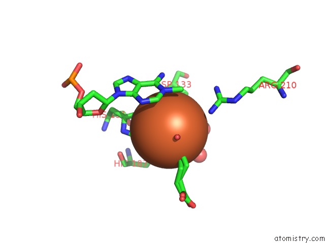
Mono view
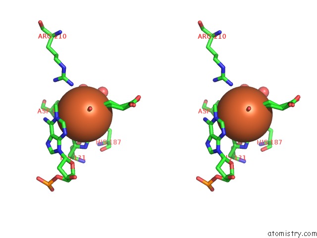
Stereo pair view
Mono view
Stereo pair view
A full contact list of Iron with other atoms in the Fe binding
site number 1 of Crystal Structure of Alkb in Complex with Fe(II), 2-Oxoglutarate, and Methylated Trinucleotide T-Mea-T (Air 3 Hours) within 5.0Å range:
|
Reference:
B.Yu,
W.C.Edstrom,
J.Benach,
Y.Hamuro,
P.C.Weber,
B.R.Gibney,
J.F.Hunt.
Crystal Structures of Catalytic Complexes of the Oxidative Dna/Rna Repair Enzyme Alkb. Nature V. 439 879 2006.
ISSN: ISSN 0028-0836
PubMed: 16482161
DOI: 10.1038/NATURE04561
Page generated: Sat Aug 3 21:17:23 2024
ISSN: ISSN 0028-0836
PubMed: 16482161
DOI: 10.1038/NATURE04561
Last articles
Zn in 9MJ5Zn in 9HNW
Zn in 9G0L
Zn in 9FNE
Zn in 9DZN
Zn in 9E0I
Zn in 9D32
Zn in 9DAK
Zn in 8ZXC
Zn in 8ZUF