Iron »
PDB 2g6o-2grx »
2gbx »
Iron in PDB 2gbx: Crystal Structure of Biphenyl 2,3-Dioxygenase From Sphingomonas Yanoikuyae B1 Bound to Biphenyl
Protein crystallography data
The structure of Crystal Structure of Biphenyl 2,3-Dioxygenase From Sphingomonas Yanoikuyae B1 Bound to Biphenyl, PDB code: 2gbx
was solved by
D.J.Ferraro,
E.N.Brown,
C.Yu,
R.E.Parales,
D.T.Gibson,
S.Ramaswamy,
with X-Ray Crystallography technique. A brief refinement statistics is given in the table below:
| Resolution Low / High (Å) | 19.48 / 2.80 |
| Space group | P 31 2 1 |
| Cell size a, b, c (Å), α, β, γ (°) | 133.940, 133.940, 219.708, 90.00, 90.00, 120.00 |
| R / Rfree (%) | 23.7 / 26.8 |
Other elements in 2gbx:
The structure of Crystal Structure of Biphenyl 2,3-Dioxygenase From Sphingomonas Yanoikuyae B1 Bound to Biphenyl also contains other interesting chemical elements:
| Zinc | (Zn) | 9 atoms |
Iron Binding Sites:
The binding sites of Iron atom in the Crystal Structure of Biphenyl 2,3-Dioxygenase From Sphingomonas Yanoikuyae B1 Bound to Biphenyl
(pdb code 2gbx). This binding sites where shown within
5.0 Angstroms radius around Iron atom.
In total 9 binding sites of Iron where determined in the Crystal Structure of Biphenyl 2,3-Dioxygenase From Sphingomonas Yanoikuyae B1 Bound to Biphenyl, PDB code: 2gbx:
Jump to Iron binding site number: 1; 2; 3; 4; 5; 6; 7; 8; 9;
In total 9 binding sites of Iron where determined in the Crystal Structure of Biphenyl 2,3-Dioxygenase From Sphingomonas Yanoikuyae B1 Bound to Biphenyl, PDB code: 2gbx:
Jump to Iron binding site number: 1; 2; 3; 4; 5; 6; 7; 8; 9;
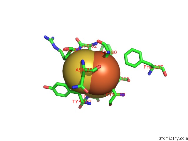
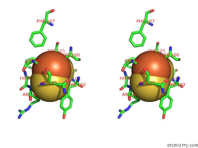
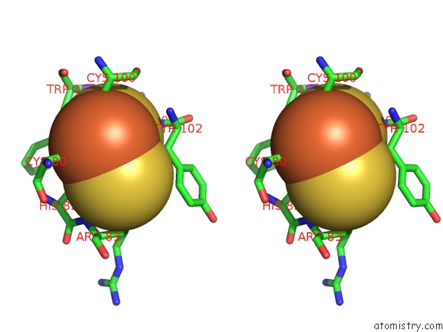
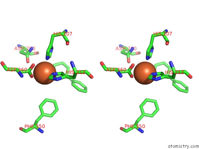
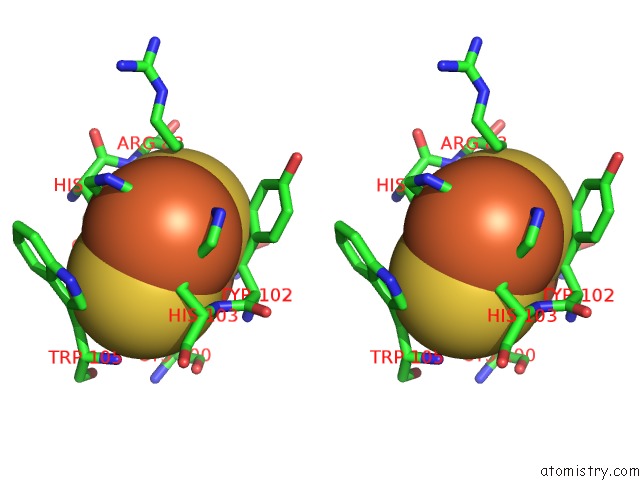
Iron binding site 1 out of 9 in 2gbx
Go back to
Iron binding site 1 out
of 9 in the Crystal Structure of Biphenyl 2,3-Dioxygenase From Sphingomonas Yanoikuyae B1 Bound to Biphenyl
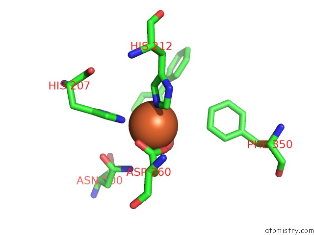
Mono view
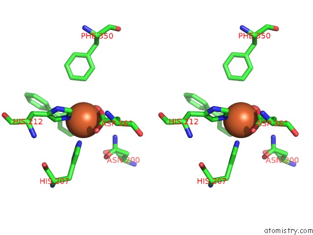
Stereo pair view
Mono view
Stereo pair view
A full contact list of Iron with other atoms in the Fe binding
site number 1 of Crystal Structure of Biphenyl 2,3-Dioxygenase From Sphingomonas Yanoikuyae B1 Bound to Biphenyl within 5.0Å range:
|
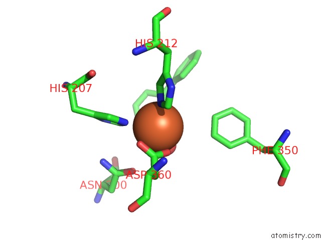
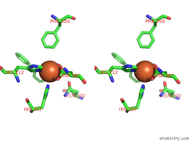
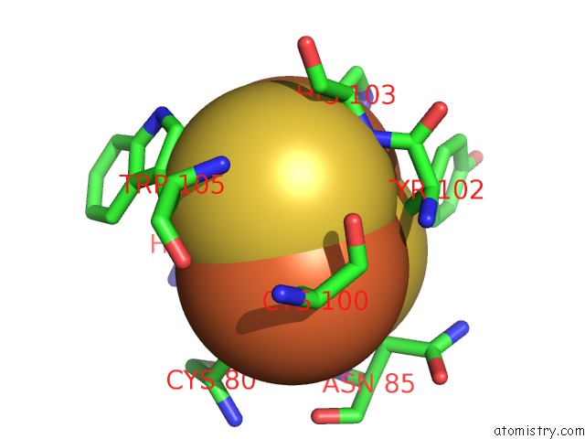
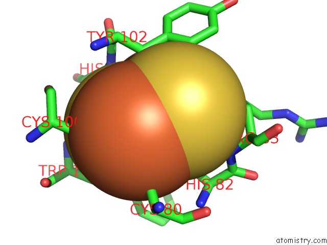
Iron binding site 2 out of 9 in 2gbx
Go back to
Iron binding site 2 out
of 9 in the Crystal Structure of Biphenyl 2,3-Dioxygenase From Sphingomonas Yanoikuyae B1 Bound to Biphenyl
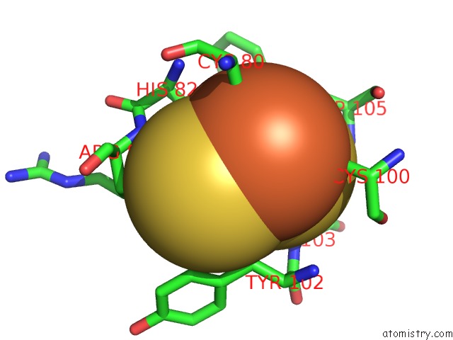
Mono view
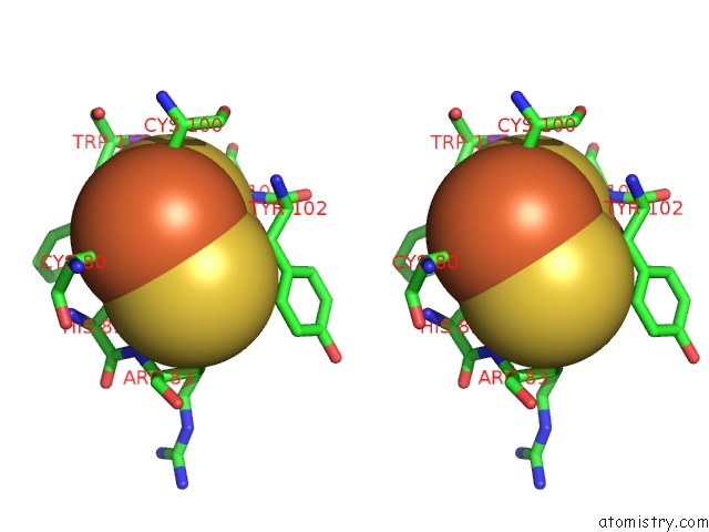
Stereo pair view
Mono view
Stereo pair view
A full contact list of Iron with other atoms in the Fe binding
site number 2 of Crystal Structure of Biphenyl 2,3-Dioxygenase From Sphingomonas Yanoikuyae B1 Bound to Biphenyl within 5.0Å range:
|
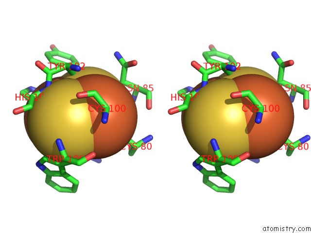
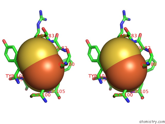
Iron binding site 3 out of 9 in 2gbx
Go back to
Iron binding site 3 out
of 9 in the Crystal Structure of Biphenyl 2,3-Dioxygenase From Sphingomonas Yanoikuyae B1 Bound to Biphenyl
Mono view
Stereo pair view
Mono view
Stereo pair view
A full contact list of Iron with other atoms in the Fe binding
site number 3 of Crystal Structure of Biphenyl 2,3-Dioxygenase From Sphingomonas Yanoikuyae B1 Bound to Biphenyl within 5.0Å range:
|
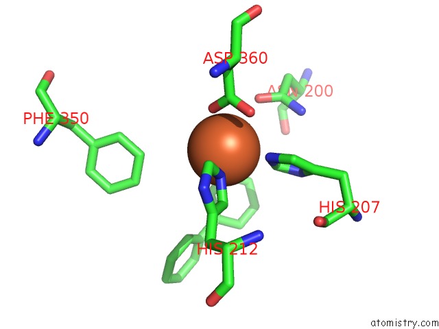
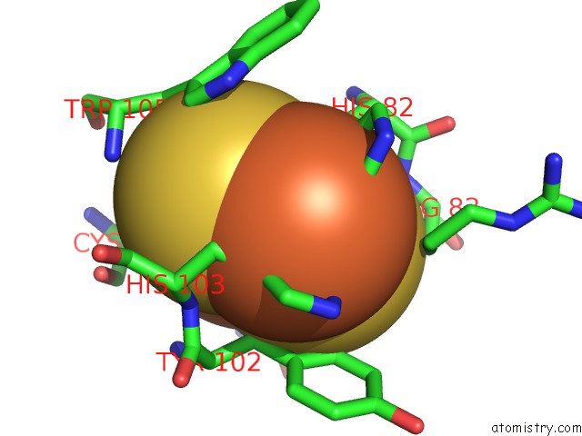
Iron binding site 4 out of 9 in 2gbx
Go back to
Iron binding site 4 out
of 9 in the Crystal Structure of Biphenyl 2,3-Dioxygenase From Sphingomonas Yanoikuyae B1 Bound to Biphenyl
Mono view
Stereo pair view
Mono view
Stereo pair view
A full contact list of Iron with other atoms in the Fe binding
site number 4 of Crystal Structure of Biphenyl 2,3-Dioxygenase From Sphingomonas Yanoikuyae B1 Bound to Biphenyl within 5.0Å range:
|
Iron binding site 5 out of 9 in 2gbx
Go back to
Iron binding site 5 out
of 9 in the Crystal Structure of Biphenyl 2,3-Dioxygenase From Sphingomonas Yanoikuyae B1 Bound to Biphenyl

Mono view
Stereo pair view
Mono view
Stereo pair view
A full contact list of Iron with other atoms in the Fe binding
site number 5 of Crystal Structure of Biphenyl 2,3-Dioxygenase From Sphingomonas Yanoikuyae B1 Bound to Biphenyl within 5.0Å range:
|
Iron binding site 6 out of 9 in 2gbx
Go back to
Iron binding site 6 out
of 9 in the Crystal Structure of Biphenyl 2,3-Dioxygenase From Sphingomonas Yanoikuyae B1 Bound to Biphenyl
Mono view
Stereo pair view
Mono view
Stereo pair view
A full contact list of Iron with other atoms in the Fe binding
site number 6 of Crystal Structure of Biphenyl 2,3-Dioxygenase From Sphingomonas Yanoikuyae B1 Bound to Biphenyl within 5.0Å range:
|
Iron binding site 7 out of 9 in 2gbx
Go back to
Iron binding site 7 out
of 9 in the Crystal Structure of Biphenyl 2,3-Dioxygenase From Sphingomonas Yanoikuyae B1 Bound to Biphenyl
Mono view
Stereo pair view
Mono view
Stereo pair view
A full contact list of Iron with other atoms in the Fe binding
site number 7 of Crystal Structure of Biphenyl 2,3-Dioxygenase From Sphingomonas Yanoikuyae B1 Bound to Biphenyl within 5.0Å range:
|
Iron binding site 8 out of 9 in 2gbx
Go back to
Iron binding site 8 out
of 9 in the Crystal Structure of Biphenyl 2,3-Dioxygenase From Sphingomonas Yanoikuyae B1 Bound to Biphenyl
Mono view
Stereo pair view
Mono view
Stereo pair view
A full contact list of Iron with other atoms in the Fe binding
site number 8 of Crystal Structure of Biphenyl 2,3-Dioxygenase From Sphingomonas Yanoikuyae B1 Bound to Biphenyl within 5.0Å range:
|
Iron binding site 9 out of 9 in 2gbx
Go back to
Iron binding site 9 out
of 9 in the Crystal Structure of Biphenyl 2,3-Dioxygenase From Sphingomonas Yanoikuyae B1 Bound to Biphenyl
Mono view
Stereo pair view
Mono view
Stereo pair view
A full contact list of Iron with other atoms in the Fe binding
site number 9 of Crystal Structure of Biphenyl 2,3-Dioxygenase From Sphingomonas Yanoikuyae B1 Bound to Biphenyl within 5.0Å range:
|
Reference:
D.J.Ferraro,
E.N.Brown,
C.L.Yu,
R.E.Parales,
D.T.Gibson,
S.Ramaswamy.
Structural Investigations of the Ferredoxin and Terminal Oxygenase Components of the Biphenyl 2,3-Dioxygenase From Sphingobium Yanoikuyae B1. Bmc Struct.Biol. V. 7 10 2007.
ISSN: ESSN 1472-6807
PubMed: 17349044
DOI: 10.1186/1472-6807-7-10
Page generated: Sat Aug 3 22:30:04 2024
ISSN: ESSN 1472-6807
PubMed: 17349044
DOI: 10.1186/1472-6807-7-10
Last articles
Zn in 9MJ5Zn in 9HNW
Zn in 9G0L
Zn in 9FNE
Zn in 9DZN
Zn in 9E0I
Zn in 9D32
Zn in 9DAK
Zn in 8ZXC
Zn in 8ZUF