Iron »
PDB 2g6o-2grx »
2gok »
Iron in PDB 2gok: Crystal Structure of the Imidazolonepropionase From Agrobacterium Tumefaciens at 1.87 A Resolution
Enzymatic activity of Crystal Structure of the Imidazolonepropionase From Agrobacterium Tumefaciens at 1.87 A Resolution
All present enzymatic activity of Crystal Structure of the Imidazolonepropionase From Agrobacterium Tumefaciens at 1.87 A Resolution:
3.5.2.7;
3.5.2.7;
Protein crystallography data
The structure of Crystal Structure of the Imidazolonepropionase From Agrobacterium Tumefaciens at 1.87 A Resolution, PDB code: 2gok
was solved by
R.Tyagi,
D.Kumaran,
S.Swaminathan,
S.K.Burley,
New York Sgx Researchcenter For Structural Genomics (Nysgxrc),
with X-Ray Crystallography technique. A brief refinement statistics is given in the table below:
| Resolution Low / High (Å) | 23.64 / 1.87 |
| Space group | C 1 2 1 |
| Cell size a, b, c (Å), α, β, γ (°) | 140.754, 63.630, 103.716, 90.00, 111.93, 90.00 |
| R / Rfree (%) | 20.7 / 23.1 |
Other elements in 2gok:
The structure of Crystal Structure of the Imidazolonepropionase From Agrobacterium Tumefaciens at 1.87 A Resolution also contains other interesting chemical elements:
| Magnesium | (Mg) | 2 atoms |
| Chlorine | (Cl) | 1 atom |
Iron Binding Sites:
The binding sites of Iron atom in the Crystal Structure of the Imidazolonepropionase From Agrobacterium Tumefaciens at 1.87 A Resolution
(pdb code 2gok). This binding sites where shown within
5.0 Angstroms radius around Iron atom.
In total 2 binding sites of Iron where determined in the Crystal Structure of the Imidazolonepropionase From Agrobacterium Tumefaciens at 1.87 A Resolution, PDB code: 2gok:
Jump to Iron binding site number: 1; 2;
In total 2 binding sites of Iron where determined in the Crystal Structure of the Imidazolonepropionase From Agrobacterium Tumefaciens at 1.87 A Resolution, PDB code: 2gok:
Jump to Iron binding site number: 1; 2;
Iron binding site 1 out of 2 in 2gok
Go back to
Iron binding site 1 out
of 2 in the Crystal Structure of the Imidazolonepropionase From Agrobacterium Tumefaciens at 1.87 A Resolution
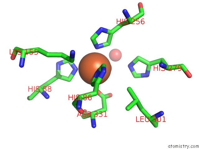
Mono view
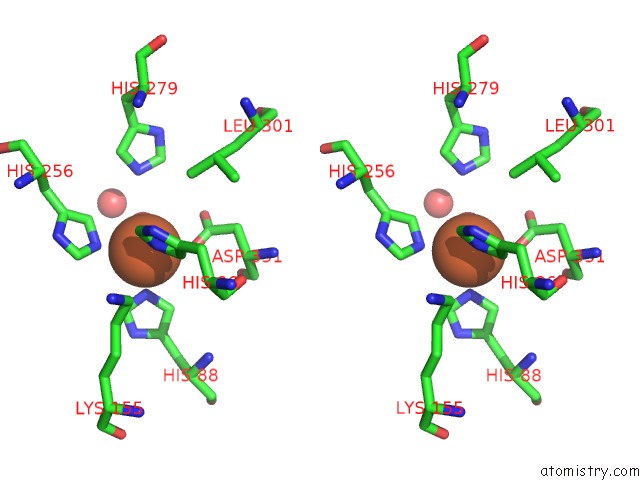
Stereo pair view
Mono view
Stereo pair view
A full contact list of Iron with other atoms in the Fe binding
site number 1 of Crystal Structure of the Imidazolonepropionase From Agrobacterium Tumefaciens at 1.87 A Resolution within 5.0Å range:
|
Iron binding site 2 out of 2 in 2gok
Go back to
Iron binding site 2 out
of 2 in the Crystal Structure of the Imidazolonepropionase From Agrobacterium Tumefaciens at 1.87 A Resolution
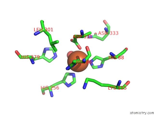
Mono view
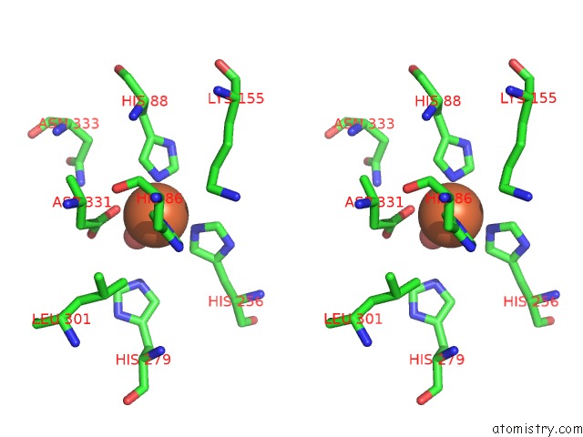
Stereo pair view
Mono view
Stereo pair view
A full contact list of Iron with other atoms in the Fe binding
site number 2 of Crystal Structure of the Imidazolonepropionase From Agrobacterium Tumefaciens at 1.87 A Resolution within 5.0Å range:
|
Reference:
R.Tyagi,
D.Kumaran,
S.K.Burley,
S.Swaminathan.
X-Ray Structure of Imidazolonepropionase From Agrobacterium Tumefaciens at 1.87 A Resolution. Proteins V. 69 652 2007.
ISSN: ISSN 0887-3585
PubMed: 17640072
DOI: 10.1002/PROT.21559
Page generated: Sat Aug 3 22:36:43 2024
ISSN: ISSN 0887-3585
PubMed: 17640072
DOI: 10.1002/PROT.21559
Last articles
Zn in 9MJ5Zn in 9HNW
Zn in 9G0L
Zn in 9FNE
Zn in 9DZN
Zn in 9E0I
Zn in 9D32
Zn in 9DAK
Zn in 8ZXC
Zn in 8ZUF