Iron »
PDB 2g6o-2grx »
2grh »
Iron in PDB 2grh: M37V Mutant of Scapharca Dimeric Hemoglobin, with Co Bound
Protein crystallography data
The structure of M37V Mutant of Scapharca Dimeric Hemoglobin, with Co Bound, PDB code: 2grh
was solved by
J.E.Knapp,
R.Pahl,
V.Srajer,
W.E.Royer Jr.,
with X-Ray Crystallography technique. A brief refinement statistics is given in the table below:
| Resolution Low / High (Å) | 30.61 / 1.50 |
| Space group | C 1 2 1 |
| Cell size a, b, c (Å), α, β, γ (°) | 93.040, 43.930, 83.440, 90.00, 121.95, 90.00 |
| R / Rfree (%) | 18.6 / 19.4 |
Iron Binding Sites:
The binding sites of Iron atom in the M37V Mutant of Scapharca Dimeric Hemoglobin, with Co Bound
(pdb code 2grh). This binding sites where shown within
5.0 Angstroms radius around Iron atom.
In total 2 binding sites of Iron where determined in the M37V Mutant of Scapharca Dimeric Hemoglobin, with Co Bound, PDB code: 2grh:
Jump to Iron binding site number: 1; 2;
In total 2 binding sites of Iron where determined in the M37V Mutant of Scapharca Dimeric Hemoglobin, with Co Bound, PDB code: 2grh:
Jump to Iron binding site number: 1; 2;
Iron binding site 1 out of 2 in 2grh
Go back to
Iron binding site 1 out
of 2 in the M37V Mutant of Scapharca Dimeric Hemoglobin, with Co Bound
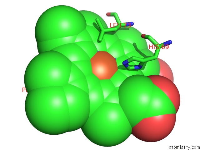
Mono view
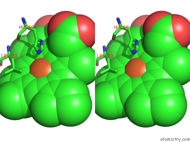
Stereo pair view
Mono view
Stereo pair view
A full contact list of Iron with other atoms in the Fe binding
site number 1 of M37V Mutant of Scapharca Dimeric Hemoglobin, with Co Bound within 5.0Å range:
|
Iron binding site 2 out of 2 in 2grh
Go back to
Iron binding site 2 out
of 2 in the M37V Mutant of Scapharca Dimeric Hemoglobin, with Co Bound
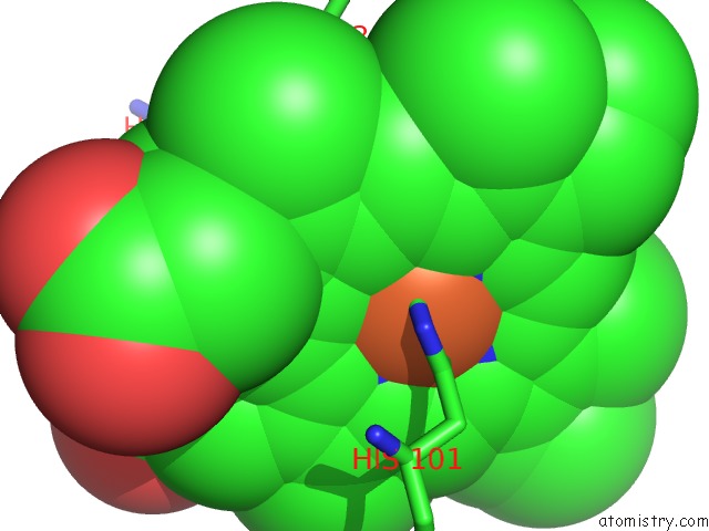
Mono view
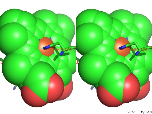
Stereo pair view
Mono view
Stereo pair view
A full contact list of Iron with other atoms in the Fe binding
site number 2 of M37V Mutant of Scapharca Dimeric Hemoglobin, with Co Bound within 5.0Å range:
|
Reference:
J.E.Knapp,
R.Pahl,
V.Srajer,
W.E.Royer Jr..
Allosteric Action in Real Time: Time-Resolved Crystallographic Studies of A Cooperative Dimeric Hemoglobin. Proc.Natl.Acad.Sci.Usa V. 103 7649 2006.
ISSN: ISSN 0027-8424
PubMed: 16684887
DOI: 10.1073/PNAS.0509411103
Page generated: Sat Aug 3 22:39:07 2024
ISSN: ISSN 0027-8424
PubMed: 16684887
DOI: 10.1073/PNAS.0509411103
Last articles
Zn in 9MJ5Zn in 9HNW
Zn in 9G0L
Zn in 9FNE
Zn in 9DZN
Zn in 9E0I
Zn in 9D32
Zn in 9DAK
Zn in 8ZXC
Zn in 8ZUF