Iron »
PDB 2grz-2hk6 »
2hhb »
Iron in PDB 2hhb: The Crystal Structure of Human Deoxyhaemoglobin at 1.74 Angstroms Resolution
Protein crystallography data
The structure of The Crystal Structure of Human Deoxyhaemoglobin at 1.74 Angstroms Resolution, PDB code: 2hhb
was solved by
G.Fermi,
M.F.Perutz,
with X-Ray Crystallography technique. A brief refinement statistics is given in the table below:
| Resolution Low / High (Å) | N/A / 1.74 |
| Space group | P 1 21 1 |
| Cell size a, b, c (Å), α, β, γ (°) | 63.150, 83.590, 53.800, 90.00, 99.34, 90.00 |
| R / Rfree (%) | 16 / n/a |
Iron Binding Sites:
The binding sites of Iron atom in the The Crystal Structure of Human Deoxyhaemoglobin at 1.74 Angstroms Resolution
(pdb code 2hhb). This binding sites where shown within
5.0 Angstroms radius around Iron atom.
In total 4 binding sites of Iron where determined in the The Crystal Structure of Human Deoxyhaemoglobin at 1.74 Angstroms Resolution, PDB code: 2hhb:
Jump to Iron binding site number: 1; 2; 3; 4;
In total 4 binding sites of Iron where determined in the The Crystal Structure of Human Deoxyhaemoglobin at 1.74 Angstroms Resolution, PDB code: 2hhb:
Jump to Iron binding site number: 1; 2; 3; 4;
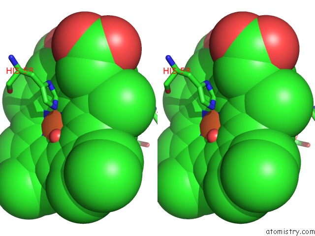
Iron binding site 1 out of 4 in 2hhb
Go back to
Iron binding site 1 out
of 4 in the The Crystal Structure of Human Deoxyhaemoglobin at 1.74 Angstroms Resolution
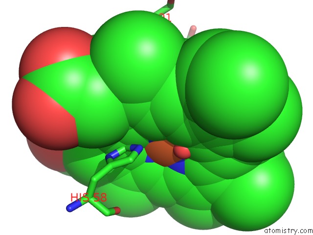
Mono view

Stereo pair view
Mono view
Stereo pair view
A full contact list of Iron with other atoms in the Fe binding
site number 1 of The Crystal Structure of Human Deoxyhaemoglobin at 1.74 Angstroms Resolution within 5.0Å range:
|
Iron binding site 2 out of 4 in 2hhb
Go back to
Iron binding site 2 out
of 4 in the The Crystal Structure of Human Deoxyhaemoglobin at 1.74 Angstroms Resolution
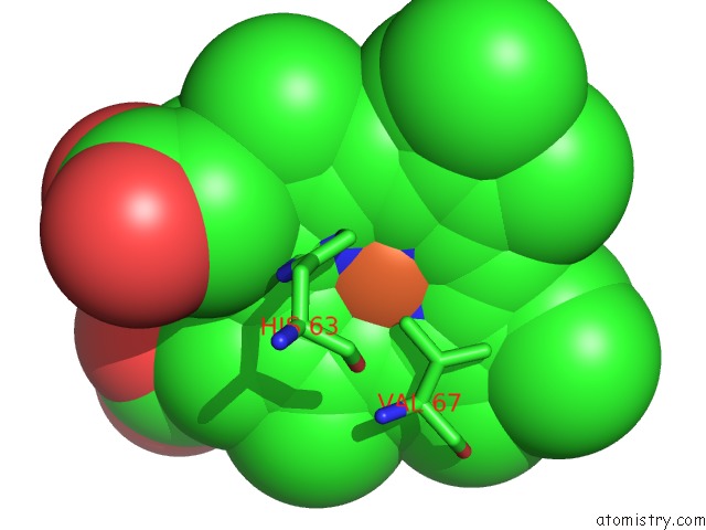
Mono view
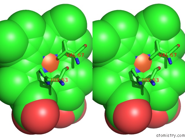
Stereo pair view
Mono view
Stereo pair view
A full contact list of Iron with other atoms in the Fe binding
site number 2 of The Crystal Structure of Human Deoxyhaemoglobin at 1.74 Angstroms Resolution within 5.0Å range:
|
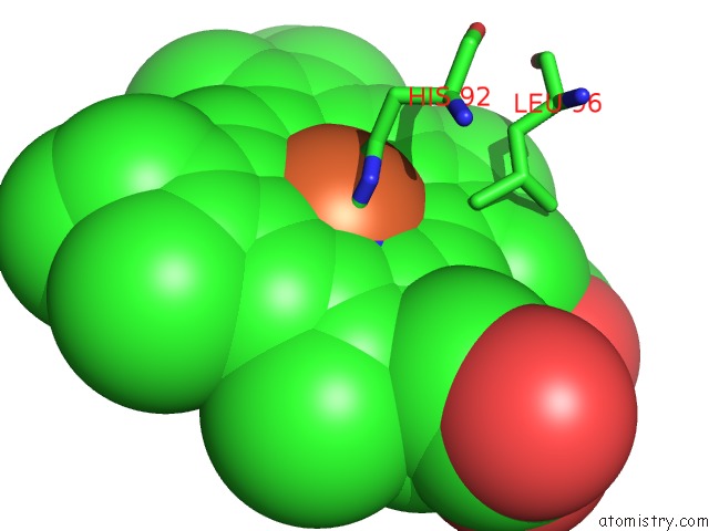
Iron binding site 3 out of 4 in 2hhb
Go back to
Iron binding site 3 out
of 4 in the The Crystal Structure of Human Deoxyhaemoglobin at 1.74 Angstroms Resolution
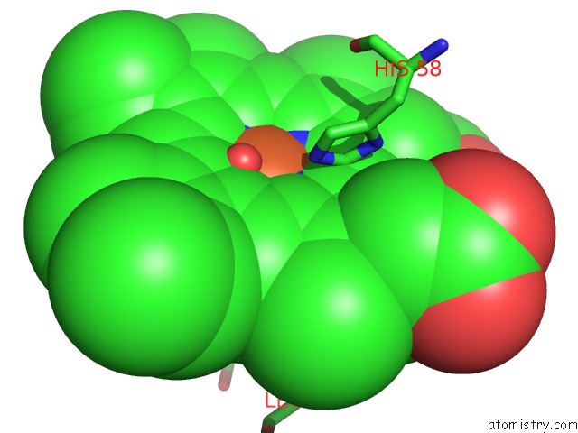
Mono view
Stereo pair view
Mono view
Stereo pair view
A full contact list of Iron with other atoms in the Fe binding
site number 3 of The Crystal Structure of Human Deoxyhaemoglobin at 1.74 Angstroms Resolution within 5.0Å range:
|
Iron binding site 4 out of 4 in 2hhb
Go back to
Iron binding site 4 out
of 4 in the The Crystal Structure of Human Deoxyhaemoglobin at 1.74 Angstroms Resolution
Mono view

Stereo pair view
Mono view
Stereo pair view
A full contact list of Iron with other atoms in the Fe binding
site number 4 of The Crystal Structure of Human Deoxyhaemoglobin at 1.74 Angstroms Resolution within 5.0Å range:
|
Reference:
G.Fermi,
M.F.Perutz,
B.Shaanan,
R.Fourme.
The Crystal Structure of Human Deoxyhaemoglobin at 1.74 A Resolution J.Mol.Biol. V. 175 159 1984.
ISSN: ISSN 0022-2836
PubMed: 6726807
DOI: 10.1016/0022-2836(84)90472-8
Page generated: Sat Aug 3 22:56:43 2024
ISSN: ISSN 0022-2836
PubMed: 6726807
DOI: 10.1016/0022-2836(84)90472-8
Last articles
Cl in 8A0OCl in 7ZYW
Cl in 7ZY8
Cl in 7ZYQ
Cl in 7ZYN
Cl in 7ZY3
Cl in 7ZYM
Cl in 7ZYK
Cl in 7ZYD
Cl in 7ZXV