Iron »
PDB 2hkx-2ibn »
2hkx »
Iron in PDB 2hkx: Structure of Cooa Mutant (N127L/S128L) From Carboxydothermus Hydrogenoformans
Protein crystallography data
The structure of Structure of Cooa Mutant (N127L/S128L) From Carboxydothermus Hydrogenoformans, PDB code: 2hkx
was solved by
N.D.Lanz,
M.Borjigin,
H.Li,
R.L.Kerby,
T.L.Poulos,
G.P.Roberts,
with X-Ray Crystallography technique. A brief refinement statistics is given in the table below:
| Resolution Low / High (Å) | 45.75 / 2.30 |
| Space group | P 21 21 21 |
| Cell size a, b, c (Å), α, β, γ (°) | 52.115, 92.873, 95.500, 90.00, 90.00, 90.00 |
| R / Rfree (%) | 22.7 / 28.6 |
Iron Binding Sites:
The binding sites of Iron atom in the Structure of Cooa Mutant (N127L/S128L) From Carboxydothermus Hydrogenoformans
(pdb code 2hkx). This binding sites where shown within
5.0 Angstroms radius around Iron atom.
In total only one binding site of Iron was determined in the Structure of Cooa Mutant (N127L/S128L) From Carboxydothermus Hydrogenoformans, PDB code: 2hkx:
In total only one binding site of Iron was determined in the Structure of Cooa Mutant (N127L/S128L) From Carboxydothermus Hydrogenoformans, PDB code: 2hkx:
Iron binding site 1 out of 1 in 2hkx
Go back to
Iron binding site 1 out
of 1 in the Structure of Cooa Mutant (N127L/S128L) From Carboxydothermus Hydrogenoformans
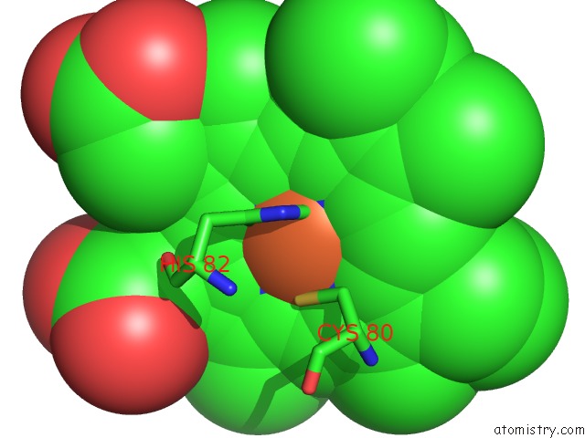
Mono view
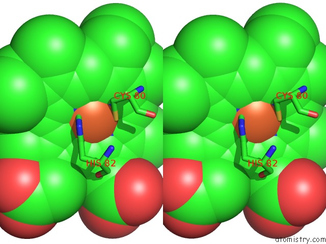
Stereo pair view
Mono view
Stereo pair view
A full contact list of Iron with other atoms in the Fe binding
site number 1 of Structure of Cooa Mutant (N127L/S128L) From Carboxydothermus Hydrogenoformans within 5.0Å range:
|
Reference:
M.Borjigin,
H.Li,
N.D.Lanz,
R.L.Kerby,
G.P.Roberts,
T.L.Poulos.
Structure-Based Hypothesis on the Activation of the Co-Sensing Transcription Factor Cooa. Acta Crystallogr.,Sect.D V. 63 282 2007.
ISSN: ISSN 0907-4449
PubMed: 17327664
DOI: 10.1107/S0907444906051638
Page generated: Sat Aug 3 23:04:40 2024
ISSN: ISSN 0907-4449
PubMed: 17327664
DOI: 10.1107/S0907444906051638
Last articles
Zn in 9MJ5Zn in 9HNW
Zn in 9G0L
Zn in 9FNE
Zn in 9DZN
Zn in 9E0I
Zn in 9D32
Zn in 9DAK
Zn in 8ZXC
Zn in 8ZUF