Iron »
PDB 2hkx-2ibn »
2ht9 »
Iron in PDB 2ht9: The Structure of Dimeric Human Glutaredoxin 2
Protein crystallography data
The structure of The Structure of Dimeric Human Glutaredoxin 2, PDB code: 2ht9
was solved by
C.Johansson,
C.Smee,
K.L.Kavanagh,
J.Debreczeni,
F.Von Delft,
O.Gileadi,
C.Arrowsmith,
J.Weigelt,
A.Edwards,
M.Sundstrom,
U.Oppermann,
Structuralgenomics Consortium (Sgc),
with X-Ray Crystallography technique. A brief refinement statistics is given in the table below:
| Resolution Low / High (Å) | 96.67 / 1.90 |
| Space group | P 63 |
| Cell size a, b, c (Å), α, β, γ (°) | 111.754, 111.754, 51.647, 90.00, 90.00, 120.00 |
| R / Rfree (%) | 14.9 / 17.8 |
Iron Binding Sites:
The binding sites of Iron atom in the The Structure of Dimeric Human Glutaredoxin 2
(pdb code 2ht9). This binding sites where shown within
5.0 Angstroms radius around Iron atom.
In total 2 binding sites of Iron where determined in the The Structure of Dimeric Human Glutaredoxin 2, PDB code: 2ht9:
Jump to Iron binding site number: 1; 2;
In total 2 binding sites of Iron where determined in the The Structure of Dimeric Human Glutaredoxin 2, PDB code: 2ht9:
Jump to Iron binding site number: 1; 2;
Iron binding site 1 out of 2 in 2ht9
Go back to
Iron binding site 1 out
of 2 in the The Structure of Dimeric Human Glutaredoxin 2
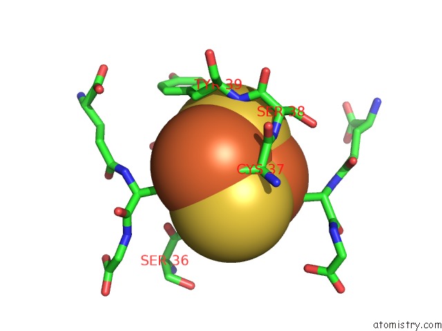
Mono view

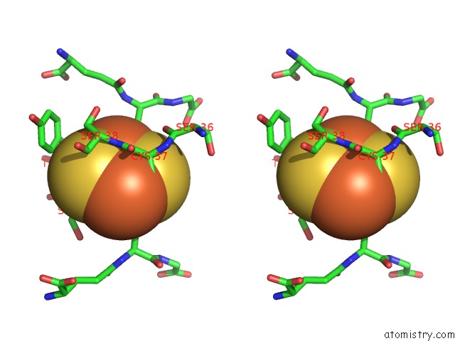
Stereo pair view
Mono view
Stereo pair view
A full contact list of Iron with other atoms in the Fe binding
site number 1 of The Structure of Dimeric Human Glutaredoxin 2 within 5.0Å range:
|
Iron binding site 2 out of 2 in 2ht9
Go back to
Iron binding site 2 out
of 2 in the The Structure of Dimeric Human Glutaredoxin 2
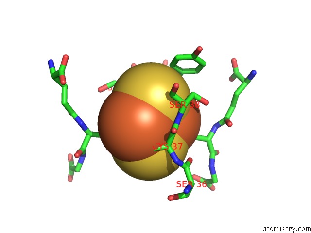
Mono view
Stereo pair view
Mono view
Stereo pair view
A full contact list of Iron with other atoms in the Fe binding
site number 2 of The Structure of Dimeric Human Glutaredoxin 2 within 5.0Å range:
|
Reference:
C.Johansson,
K.L.Kavanagh,
O.Gileadi,
U.Oppermann.
Reversible Sequestration of Active Site Cysteines in A 2FE-2S-Bridged Dimer Provides A Mechanism For Glutaredoxin 2 Regulation in Human Mitochondria J.Biol.Chem. V. 282 3077 2007.
ISSN: ISSN 0021-9258
PubMed: 17121859
DOI: 10.1074/JBC.M608179200
Page generated: Sat Aug 3 23:08:05 2024
ISSN: ISSN 0021-9258
PubMed: 17121859
DOI: 10.1074/JBC.M608179200
Last articles
Zn in 9J0NZn in 9J0O
Zn in 9J0P
Zn in 9FJX
Zn in 9EKB
Zn in 9C0F
Zn in 9CAH
Zn in 9CH0
Zn in 9CH3
Zn in 9CH1