Iron »
PDB 2hkx-2ibn »
2hys »
Iron in PDB 2hys: Crystal Structure of Nitrophorin 2 Complexed with Cyanide
Protein crystallography data
The structure of Crystal Structure of Nitrophorin 2 Complexed with Cyanide, PDB code: 2hys
was solved by
A.Weichsel,
W.R.Montfort,
with X-Ray Crystallography technique. A brief refinement statistics is given in the table below:
| Resolution Low / High (Å) | 22.00 / 1.20 |
| Space group | P 41 21 2 |
| Cell size a, b, c (Å), α, β, γ (°) | 34.377, 34.377, 257.256, 90.00, 90.00, 90.00 |
| R / Rfree (%) | 18.6 / 21.6 |
Iron Binding Sites:
The binding sites of Iron atom in the Crystal Structure of Nitrophorin 2 Complexed with Cyanide
(pdb code 2hys). This binding sites where shown within
5.0 Angstroms radius around Iron atom.
In total only one binding site of Iron was determined in the Crystal Structure of Nitrophorin 2 Complexed with Cyanide, PDB code: 2hys:
In total only one binding site of Iron was determined in the Crystal Structure of Nitrophorin 2 Complexed with Cyanide, PDB code: 2hys:
Iron binding site 1 out of 1 in 2hys
Go back to
Iron binding site 1 out
of 1 in the Crystal Structure of Nitrophorin 2 Complexed with Cyanide
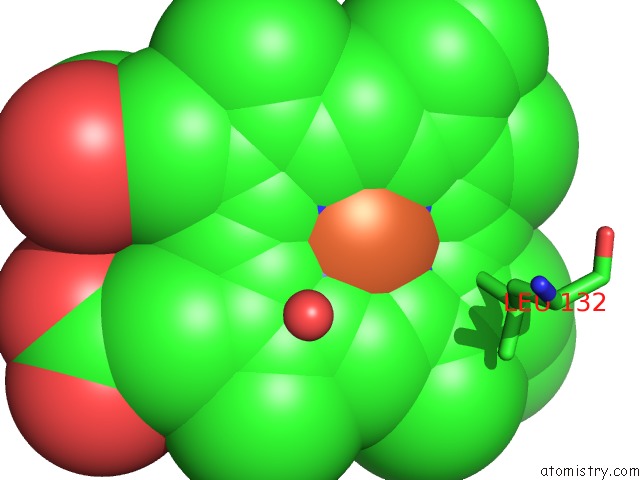
Mono view
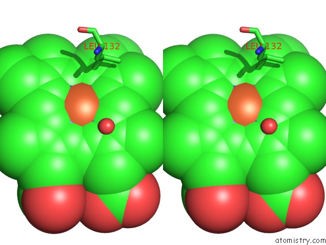
Stereo pair view
Mono view
Stereo pair view
A full contact list of Iron with other atoms in the Fe binding
site number 1 of Crystal Structure of Nitrophorin 2 Complexed with Cyanide within 5.0Å range:
|
Reference:
T.Kh.Shokhireva,
A.Weichsel,
K.M.Smith,
R.E.Berry,
N.V.Shokhirev,
C.A.Balfour,
H.Zhang,
W.R.Montfort,
F.A.Walker.
Assignment of the Ferriheme Resonances of the Low-Spin Complexes of Nitrophorins 1 and 4 By (1)H and (13)C uc(Nmr) Spectroscopy: Comparison to Structural Data Obtained From X-Ray Crystallography. Inorg.Chem. V. 46 2041 2007.
ISSN: ISSN 0020-1669
PubMed: 17290983
DOI: 10.1021/IC061408L
Page generated: Thu Jul 17 02:11:20 2025
ISSN: ISSN 0020-1669
PubMed: 17290983
DOI: 10.1021/IC061408L
Last articles
Fe in 2YXOFe in 2YRS
Fe in 2YXC
Fe in 2YNM
Fe in 2YVJ
Fe in 2YP1
Fe in 2YU2
Fe in 2YU1
Fe in 2YQB
Fe in 2YOO