Iron »
PDB 2hkx-2ibn »
2i5n »
Iron in PDB 2i5n: 1.96 A X-Ray Structure of Photosynthetic Reaction Center From Rhodopseudomonas Viridis:Crystals Grown By Microfluidic Technique
Protein crystallography data
The structure of 1.96 A X-Ray Structure of Photosynthetic Reaction Center From Rhodopseudomonas Viridis:Crystals Grown By Microfluidic Technique, PDB code: 2i5n
was solved by
L.Li,
D.Mustafi,
Q.Fu,
V.Tereshko,
D.L.Chen,
J.D.Tice,
R.F.Ismagilov,
with X-Ray Crystallography technique. A brief refinement statistics is given in the table below:
| Resolution Low / High (Å) | 20.00 / 1.96 |
| Space group | P 43 21 2 |
| Cell size a, b, c (Å), α, β, γ (°) | 220.400, 220.400, 113.009, 90.00, 90.00, 90.00 |
| R / Rfree (%) | 17.2 / 19 |
Other elements in 2i5n:
The structure of 1.96 A X-Ray Structure of Photosynthetic Reaction Center From Rhodopseudomonas Viridis:Crystals Grown By Microfluidic Technique also contains other interesting chemical elements:
| Magnesium | (Mg) | 4 atoms |
Iron Binding Sites:
The binding sites of Iron atom in the 1.96 A X-Ray Structure of Photosynthetic Reaction Center From Rhodopseudomonas Viridis:Crystals Grown By Microfluidic Technique
(pdb code 2i5n). This binding sites where shown within
5.0 Angstroms radius around Iron atom.
In total 5 binding sites of Iron where determined in the 1.96 A X-Ray Structure of Photosynthetic Reaction Center From Rhodopseudomonas Viridis:Crystals Grown By Microfluidic Technique, PDB code: 2i5n:
Jump to Iron binding site number: 1; 2; 3; 4; 5;
In total 5 binding sites of Iron where determined in the 1.96 A X-Ray Structure of Photosynthetic Reaction Center From Rhodopseudomonas Viridis:Crystals Grown By Microfluidic Technique, PDB code: 2i5n:
Jump to Iron binding site number: 1; 2; 3; 4; 5;
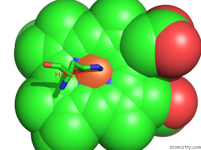
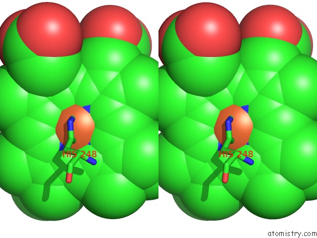
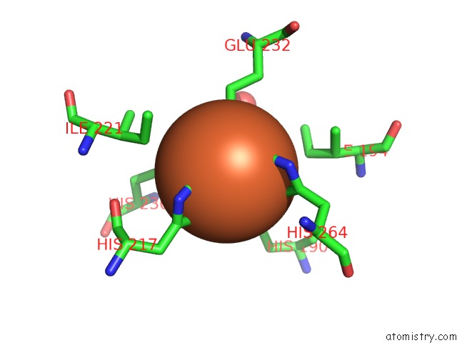
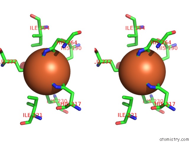
Iron binding site 1 out of 5 in 2i5n
Go back to
Iron binding site 1 out
of 5 in the 1.96 A X-Ray Structure of Photosynthetic Reaction Center From Rhodopseudomonas Viridis:Crystals Grown By Microfluidic Technique
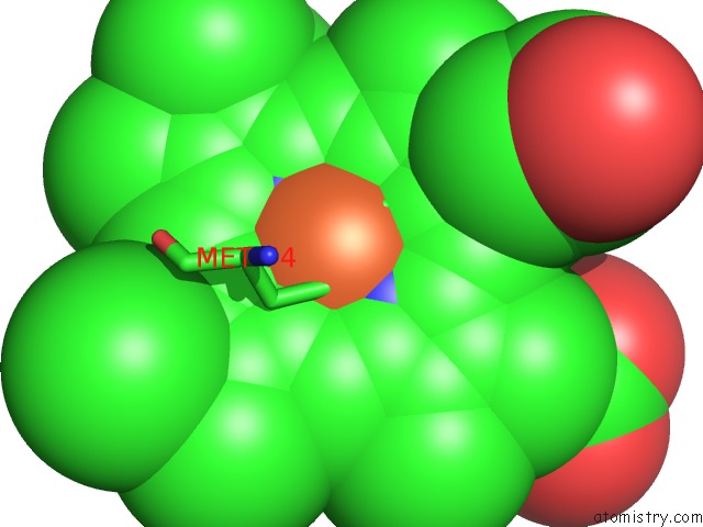
Mono view
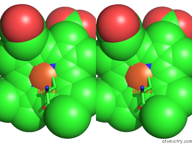
Stereo pair view
Mono view
Stereo pair view
A full contact list of Iron with other atoms in the Fe binding
site number 1 of 1.96 A X-Ray Structure of Photosynthetic Reaction Center From Rhodopseudomonas Viridis:Crystals Grown By Microfluidic Technique within 5.0Å range:
|
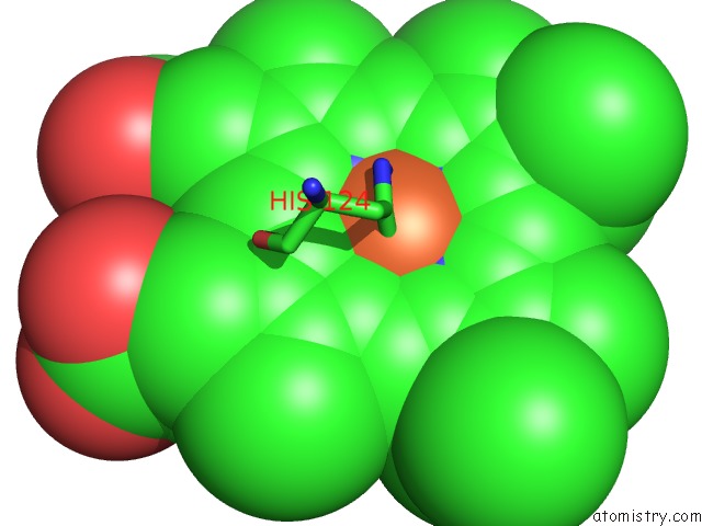
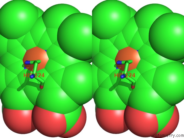
Iron binding site 2 out of 5 in 2i5n
Go back to
Iron binding site 2 out
of 5 in the 1.96 A X-Ray Structure of Photosynthetic Reaction Center From Rhodopseudomonas Viridis:Crystals Grown By Microfluidic Technique
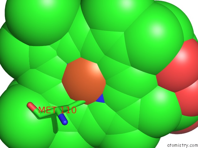
Mono view
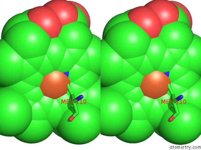
Stereo pair view
Mono view
Stereo pair view
A full contact list of Iron with other atoms in the Fe binding
site number 2 of 1.96 A X-Ray Structure of Photosynthetic Reaction Center From Rhodopseudomonas Viridis:Crystals Grown By Microfluidic Technique within 5.0Å range:
|
Iron binding site 3 out of 5 in 2i5n
Go back to
Iron binding site 3 out
of 5 in the 1.96 A X-Ray Structure of Photosynthetic Reaction Center From Rhodopseudomonas Viridis:Crystals Grown By Microfluidic Technique
Mono view
Stereo pair view
Mono view
Stereo pair view
A full contact list of Iron with other atoms in the Fe binding
site number 3 of 1.96 A X-Ray Structure of Photosynthetic Reaction Center From Rhodopseudomonas Viridis:Crystals Grown By Microfluidic Technique within 5.0Å range:
|
Iron binding site 4 out of 5 in 2i5n
Go back to
Iron binding site 4 out
of 5 in the 1.96 A X-Ray Structure of Photosynthetic Reaction Center From Rhodopseudomonas Viridis:Crystals Grown By Microfluidic Technique
Mono view
Stereo pair view
Mono view
Stereo pair view
A full contact list of Iron with other atoms in the Fe binding
site number 4 of 1.96 A X-Ray Structure of Photosynthetic Reaction Center From Rhodopseudomonas Viridis:Crystals Grown By Microfluidic Technique within 5.0Å range:
|
Iron binding site 5 out of 5 in 2i5n
Go back to
Iron binding site 5 out
of 5 in the 1.96 A X-Ray Structure of Photosynthetic Reaction Center From Rhodopseudomonas Viridis:Crystals Grown By Microfluidic Technique
Mono view
Stereo pair view
Mono view
Stereo pair view
A full contact list of Iron with other atoms in the Fe binding
site number 5 of 1.96 A X-Ray Structure of Photosynthetic Reaction Center From Rhodopseudomonas Viridis:Crystals Grown By Microfluidic Technique within 5.0Å range:
|
Reference:
L.Li,
D.Mustafi,
Q.Fu,
V.Tereshko,
D.L.Chen,
J.D.Tice,
R.F.Ismagilov.
Nanoliter Microfluidic Hybrid Method For Simultaneous Screening and Optimization Validated with Crystallization of Membrane Proteins. Proc.Natl.Acad.Sci.Usa V. 103 19243 2006.
ISSN: ISSN 0027-8424
PubMed: 17159147
DOI: 10.1073/PNAS.0607502103
Page generated: Sat Aug 3 23:13:05 2024
ISSN: ISSN 0027-8424
PubMed: 17159147
DOI: 10.1073/PNAS.0607502103
Last articles
Zn in 9MJ5Zn in 9HNW
Zn in 9G0L
Zn in 9FNE
Zn in 9DZN
Zn in 9E0I
Zn in 9D32
Zn in 9DAK
Zn in 8ZXC
Zn in 8ZUF