Iron »
PDB 2hkx-2ibn »
2ia8 »
Iron in PDB 2ia8: Kinetic and Crystallographic Studies of A Redesigned Manganese-Binding Site in Cytochrome C Peroxidase
Enzymatic activity of Kinetic and Crystallographic Studies of A Redesigned Manganese-Binding Site in Cytochrome C Peroxidase
All present enzymatic activity of Kinetic and Crystallographic Studies of A Redesigned Manganese-Binding Site in Cytochrome C Peroxidase:
1.11.1.5;
1.11.1.5;
Protein crystallography data
The structure of Kinetic and Crystallographic Studies of A Redesigned Manganese-Binding Site in Cytochrome C Peroxidase, PDB code: 2ia8
was solved by
T.Pfister,
A.Y.Mirarefi,
A.J.Gengenbach,
X.Zhao,
C.D.N.Conaster,
Y.G.Gao,
H.Robinson,
C.F.Zukoski,
A.H.J.Wang,
Y.Lu,
with X-Ray Crystallography technique. A brief refinement statistics is given in the table below:
| Resolution Low / High (Å) | 10.00 / 1.48 |
| Space group | P 21 21 21 |
| Cell size a, b, c (Å), α, β, γ (°) | 44.192, 52.612, 136.137, 90.00, 90.00, 90.00 |
| R / Rfree (%) | 21.2 / 24.7 |
Iron Binding Sites:
The binding sites of Iron atom in the Kinetic and Crystallographic Studies of A Redesigned Manganese-Binding Site in Cytochrome C Peroxidase
(pdb code 2ia8). This binding sites where shown within
5.0 Angstroms radius around Iron atom.
In total only one binding site of Iron was determined in the Kinetic and Crystallographic Studies of A Redesigned Manganese-Binding Site in Cytochrome C Peroxidase, PDB code: 2ia8:
In total only one binding site of Iron was determined in the Kinetic and Crystallographic Studies of A Redesigned Manganese-Binding Site in Cytochrome C Peroxidase, PDB code: 2ia8:
Iron binding site 1 out of 1 in 2ia8
Go back to
Iron binding site 1 out
of 1 in the Kinetic and Crystallographic Studies of A Redesigned Manganese-Binding Site in Cytochrome C Peroxidase
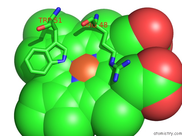
Mono view
Stereo pair view
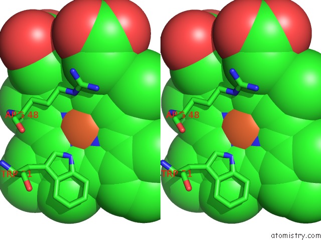
Mono view
Stereo pair view
A full contact list of Iron with other atoms in the Fe binding
site number 1 of Kinetic and Crystallographic Studies of A Redesigned Manganese-Binding Site in Cytochrome C Peroxidase within 5.0Å range:
|
Reference:
T.D.Pfister,
A.Y.Mirarefi,
A.J.Gengenbach,
X.Zhao,
C.Danstrom,
N.Conatser,
Y.-G.Gao,
H.Robinson,
C.F.Zukoski,
A.H.-J.Wang,
Y.Lu.
Kinetic and Crystallographic Studies of A Redesigned Manganese-Binding Site in Cytochrome C Peroxidase J.Biol.Inorg.Chem. V. 12 126 2007.
ISSN: ISSN 0949-8257
PubMed: 17021923
DOI: 10.1007/S00775-006-0171-0
Page generated: Sat Aug 3 23:14:07 2024
ISSN: ISSN 0949-8257
PubMed: 17021923
DOI: 10.1007/S00775-006-0171-0
Last articles
Zn in 9MJ5Zn in 9HNW
Zn in 9G0L
Zn in 9FNE
Zn in 9DZN
Zn in 9E0I
Zn in 9D32
Zn in 9DAK
Zn in 8ZXC
Zn in 8ZUF