Iron »
PDB 2hkx-2ibn »
2ibn »
Iron in PDB 2ibn: Crystal Structure of Human Myo-Inositol Oxygenase (Miox)
Enzymatic activity of Crystal Structure of Human Myo-Inositol Oxygenase (Miox)
All present enzymatic activity of Crystal Structure of Human Myo-Inositol Oxygenase (Miox):
1.13.99.1;
1.13.99.1;
Protein crystallography data
The structure of Crystal Structure of Human Myo-Inositol Oxygenase (Miox), PDB code: 2ibn
was solved by
B.M.Hallberg,
R.D.Busam,
C.Arrowsmith,
H.Berglund,
R.Collins,
A.Edwards,
M.Ehn,
S.Flodin,
A.Flores,
S.Graslund,
M.Hammarstrom,
M.Hogbom,
L.Holmberg-Schiavone,
I.Johansson,
T.Karlberg,
T.Kotenyova,
P.Nilsson-Ehle,
P.Nordlund,
T.Nyman,
D.Ogg,
J.Sagemark,
P.Stenmark,
M.Sundstrom,
J.Uppenberg,
S.Van Den Berg,
J.Weigelt,
A.G.Thorsell,
C.Persson,
Structural Genomics Consortium (Sgc),
with X-Ray Crystallography technique. A brief refinement statistics is given in the table below:
| Resolution Low / High (Å) | 29.34 / 1.50 |
| Space group | C 1 2 1 |
| Cell size a, b, c (Å), α, β, γ (°) | 119.715, 55.861, 111.516, 90.00, 116.72, 90.00 |
| R / Rfree (%) | 20.6 / 24.8 |
Iron Binding Sites:
The binding sites of Iron atom in the Crystal Structure of Human Myo-Inositol Oxygenase (Miox)
(pdb code 2ibn). This binding sites where shown within
5.0 Angstroms radius around Iron atom.
In total 4 binding sites of Iron where determined in the Crystal Structure of Human Myo-Inositol Oxygenase (Miox), PDB code: 2ibn:
Jump to Iron binding site number: 1; 2; 3; 4;
In total 4 binding sites of Iron where determined in the Crystal Structure of Human Myo-Inositol Oxygenase (Miox), PDB code: 2ibn:
Jump to Iron binding site number: 1; 2; 3; 4;
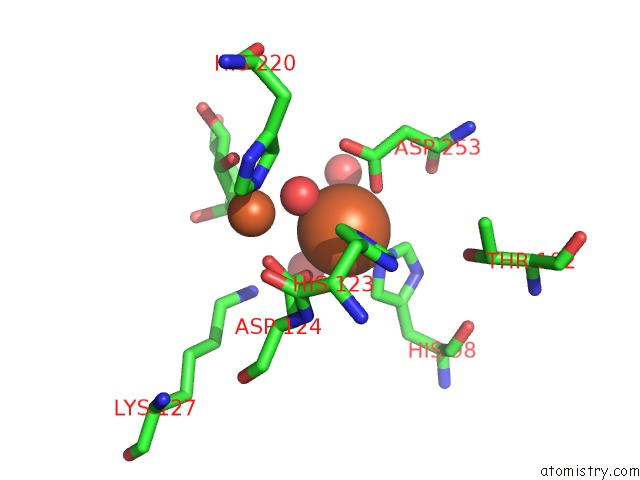
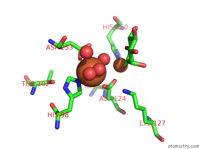
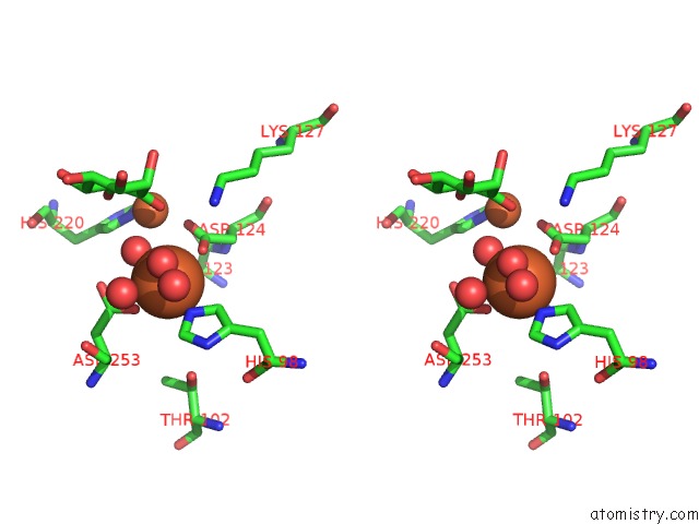
Iron binding site 1 out of 4 in 2ibn
Go back to
Iron binding site 1 out
of 4 in the Crystal Structure of Human Myo-Inositol Oxygenase (Miox)
Mono view
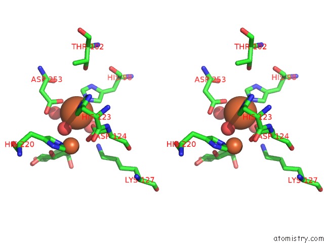
Stereo pair view
Mono view
Stereo pair view
A full contact list of Iron with other atoms in the Fe binding
site number 1 of Crystal Structure of Human Myo-Inositol Oxygenase (Miox) within 5.0Å range:
|
Iron binding site 2 out of 4 in 2ibn
Go back to
Iron binding site 2 out
of 4 in the Crystal Structure of Human Myo-Inositol Oxygenase (Miox)
Mono view
Stereo pair view
Mono view
Stereo pair view
A full contact list of Iron with other atoms in the Fe binding
site number 2 of Crystal Structure of Human Myo-Inositol Oxygenase (Miox) within 5.0Å range:
|
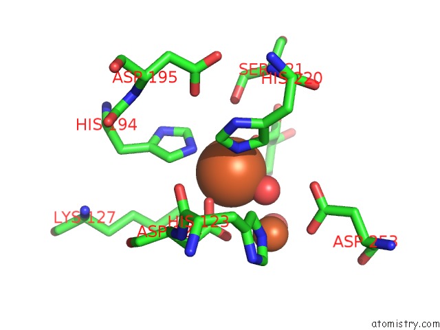
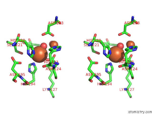
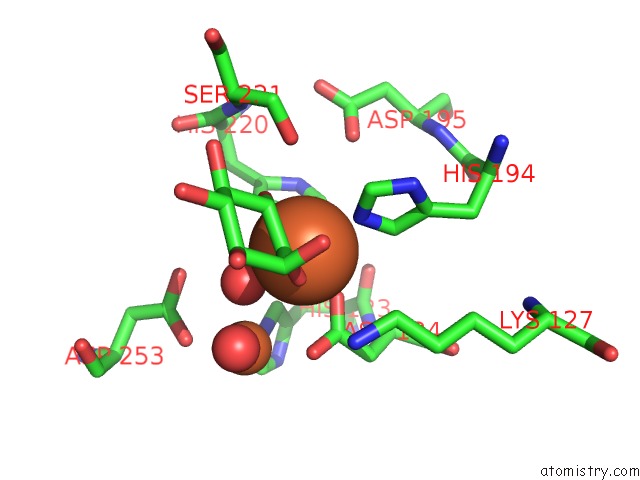
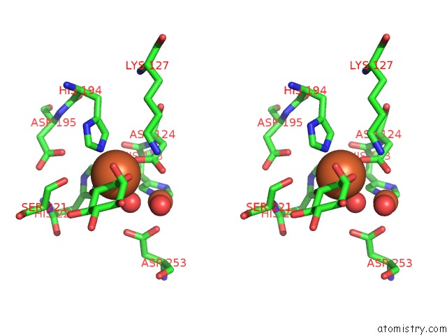
Iron binding site 3 out of 4 in 2ibn
Go back to
Iron binding site 3 out
of 4 in the Crystal Structure of Human Myo-Inositol Oxygenase (Miox)
Mono view
Stereo pair view
Mono view
Stereo pair view
A full contact list of Iron with other atoms in the Fe binding
site number 3 of Crystal Structure of Human Myo-Inositol Oxygenase (Miox) within 5.0Å range:
|
Iron binding site 4 out of 4 in 2ibn
Go back to
Iron binding site 4 out
of 4 in the Crystal Structure of Human Myo-Inositol Oxygenase (Miox)
Mono view
Stereo pair view
Mono view
Stereo pair view
A full contact list of Iron with other atoms in the Fe binding
site number 4 of Crystal Structure of Human Myo-Inositol Oxygenase (Miox) within 5.0Å range:
|
Reference:
A.G.Thorsell,
C.Persson,
N.Voevodskaya,
R.D.Busam,
M.Hammarstrom,
S.Graslund,
A.Graslund,
B.M.Hallberg.
Structural and Biophysical Characterization of Human Myo-Inositol Oxygenase J.Biol.Chem. V. 283 15209 2008.
ISSN: ISSN 0021-9258
PubMed: 18364358
DOI: 10.1074/JBC.M800348200
Page generated: Thu Jul 17 02:14:28 2025
ISSN: ISSN 0021-9258
PubMed: 18364358
DOI: 10.1074/JBC.M800348200
Last articles
Fe in 2YXOFe in 2YRS
Fe in 2YXC
Fe in 2YNM
Fe in 2YVJ
Fe in 2YP1
Fe in 2YU2
Fe in 2YU1
Fe in 2YQB
Fe in 2YOO