Iron »
PDB 2ibz-2j2f »
2iuf »
Iron in PDB 2iuf: The Structures of Penicillium Vitale Catalase: Resting State, Oxidised State (Compound I) and Complex with Aminotriazole
Enzymatic activity of The Structures of Penicillium Vitale Catalase: Resting State, Oxidised State (Compound I) and Complex with Aminotriazole
All present enzymatic activity of The Structures of Penicillium Vitale Catalase: Resting State, Oxidised State (Compound I) and Complex with Aminotriazole:
1.11.1.6;
1.11.1.6;
Protein crystallography data
The structure of The Structures of Penicillium Vitale Catalase: Resting State, Oxidised State (Compound I) and Complex with Aminotriazole, PDB code: 2iuf
was solved by
G.Murshudov,
A.Borovik,
A.Grebenko,
V.Barynin,
A.Vagin,
W.Melik-Adamyan,
with X-Ray Crystallography technique. A brief refinement statistics is given in the table below:
| Resolution Low / High (Å) | 119.52 / 1.71 |
| Space group | P 31 2 1 |
| Cell size a, b, c (Å), α, β, γ (°) | 142.440, 142.440, 132.230, 90.00, 90.00, 120.00 |
| R / Rfree (%) | 12.9 / 15.4 |
Other elements in 2iuf:
The structure of The Structures of Penicillium Vitale Catalase: Resting State, Oxidised State (Compound I) and Complex with Aminotriazole also contains other interesting chemical elements:
| Calcium | (Ca) | 6 atoms |
Iron Binding Sites:
The binding sites of Iron atom in the The Structures of Penicillium Vitale Catalase: Resting State, Oxidised State (Compound I) and Complex with Aminotriazole
(pdb code 2iuf). This binding sites where shown within
5.0 Angstroms radius around Iron atom.
In total 2 binding sites of Iron where determined in the The Structures of Penicillium Vitale Catalase: Resting State, Oxidised State (Compound I) and Complex with Aminotriazole, PDB code: 2iuf:
Jump to Iron binding site number: 1; 2;
In total 2 binding sites of Iron where determined in the The Structures of Penicillium Vitale Catalase: Resting State, Oxidised State (Compound I) and Complex with Aminotriazole, PDB code: 2iuf:
Jump to Iron binding site number: 1; 2;
Iron binding site 1 out of 2 in 2iuf
Go back to
Iron binding site 1 out
of 2 in the The Structures of Penicillium Vitale Catalase: Resting State, Oxidised State (Compound I) and Complex with Aminotriazole
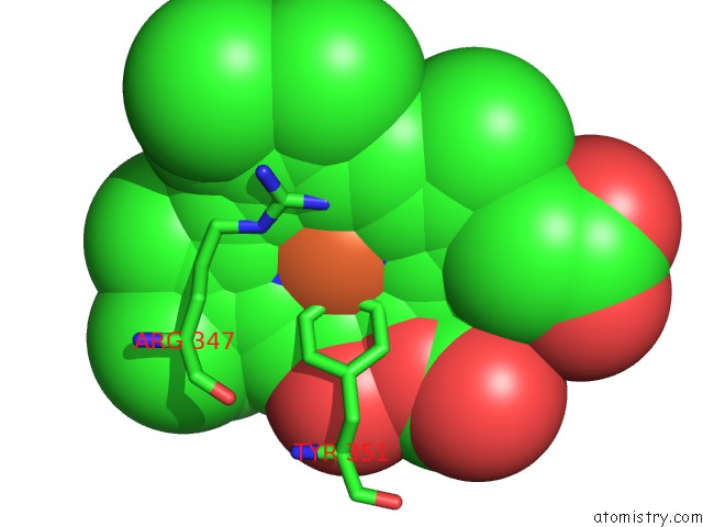
Mono view
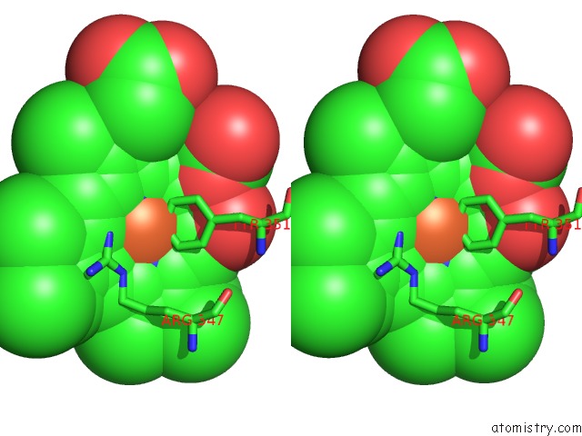
Stereo pair view
Mono view
Stereo pair view
A full contact list of Iron with other atoms in the Fe binding
site number 1 of The Structures of Penicillium Vitale Catalase: Resting State, Oxidised State (Compound I) and Complex with Aminotriazole within 5.0Å range:
|
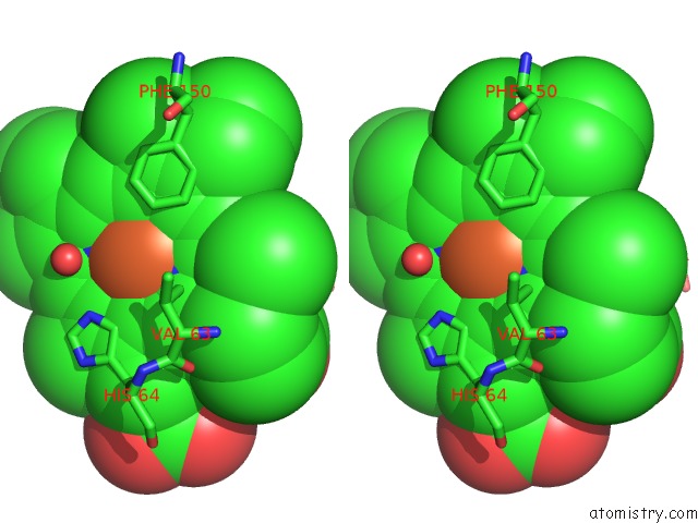
Iron binding site 2 out of 2 in 2iuf
Go back to
Iron binding site 2 out
of 2 in the The Structures of Penicillium Vitale Catalase: Resting State, Oxidised State (Compound I) and Complex with Aminotriazole
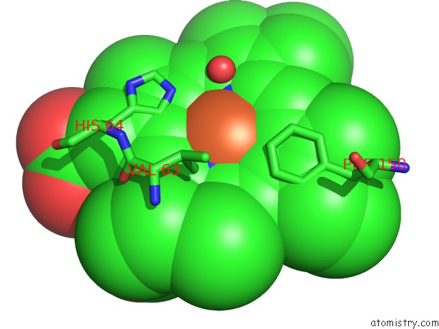
Mono view
Stereo pair view
Mono view
Stereo pair view
A full contact list of Iron with other atoms in the Fe binding
site number 2 of The Structures of Penicillium Vitale Catalase: Resting State, Oxidised State (Compound I) and Complex with Aminotriazole within 5.0Å range:
|
Reference:
M.Alfonso-Prieto,
A.Borovik,
X.Carpena,
G.Murshudov,
W.Melik-Adamyan,
I.Fita,
C.Rovira,
P.C.Loewen.
The Structures and Electronic Configuration of Compound I Intermediates of Helicobacter Pylori and Penicillium Vitale Catalases Determined By X-Ray Crystallography and Qm/Mm Density Functional Theory Calculations. J.Am.Chem.Soc. V. 129 4193 2007.
ISSN: ISSN 0002-7863
PubMed: 17358056
DOI: 10.1021/JA063660Y
Page generated: Thu Jul 17 02:19:39 2025
ISSN: ISSN 0002-7863
PubMed: 17358056
DOI: 10.1021/JA063660Y
Last articles
Fe in 2YXOFe in 2YRS
Fe in 2YXC
Fe in 2YNM
Fe in 2YVJ
Fe in 2YP1
Fe in 2YU2
Fe in 2YU1
Fe in 2YQB
Fe in 2YOO