Iron »
PDB 2j2m-2ksu »
2je3 »
Iron in PDB 2je3: Cytochrome P460 From Nitrosomonas Europaea - Probable Physiological Form
Protein crystallography data
The structure of Cytochrome P460 From Nitrosomonas Europaea - Probable Physiological Form, PDB code: 2je3
was solved by
A.R.Pearson,
B.O.Elmore,
C.Yang,
J.D.Ferrara,
A.B.Hooper,
C.M.Wilmot,
with X-Ray Crystallography technique. A brief refinement statistics is given in the table below:
| Resolution Low / High (Å) | 43.35 / 1.8 |
| Space group | P 31 2 1 |
| Cell size a, b, c (Å), α, β, γ (°) | 53.256, 53.256, 127.033, 90.00, 90.00, 120.00 |
| R / Rfree (%) | 19.5 / 23.1 |
Iron Binding Sites:
The binding sites of Iron atom in the Cytochrome P460 From Nitrosomonas Europaea - Probable Physiological Form
(pdb code 2je3). This binding sites where shown within
5.0 Angstroms radius around Iron atom.
In total only one binding site of Iron was determined in the Cytochrome P460 From Nitrosomonas Europaea - Probable Physiological Form, PDB code: 2je3:
In total only one binding site of Iron was determined in the Cytochrome P460 From Nitrosomonas Europaea - Probable Physiological Form, PDB code: 2je3:
Iron binding site 1 out of 1 in 2je3
Go back to
Iron binding site 1 out
of 1 in the Cytochrome P460 From Nitrosomonas Europaea - Probable Physiological Form
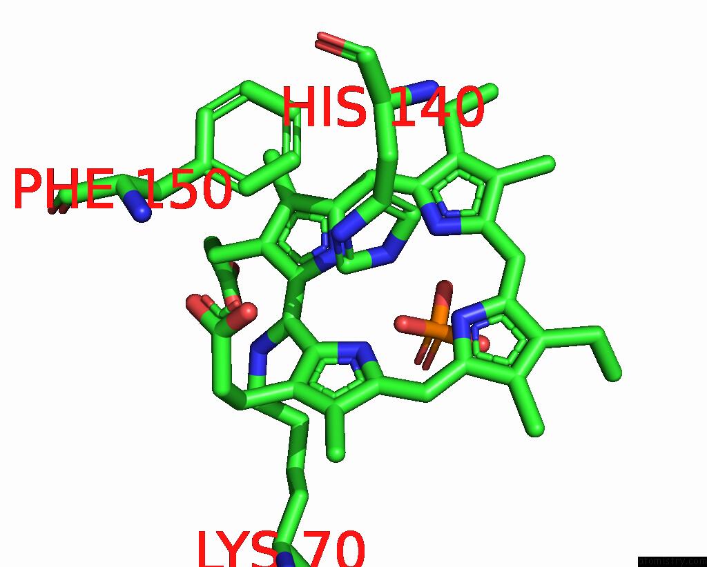
Mono view
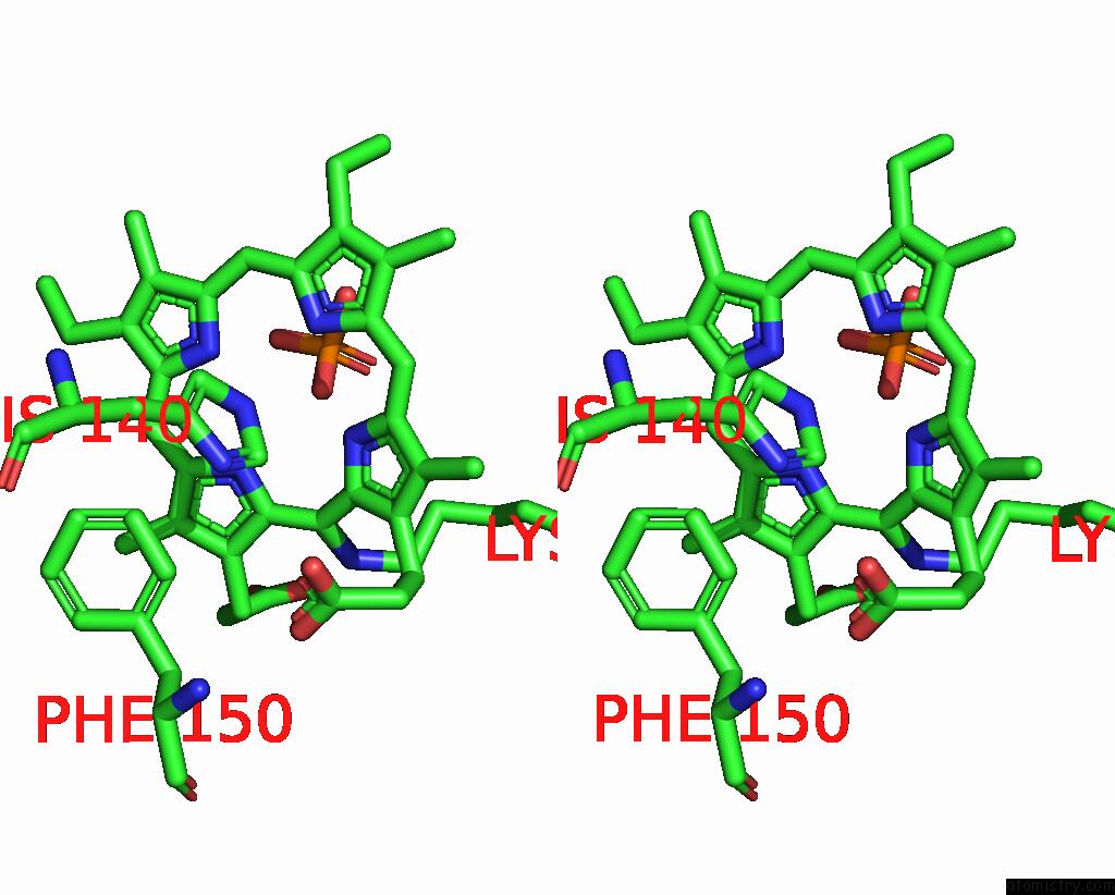
Stereo pair view
Mono view
Stereo pair view
A full contact list of Iron with other atoms in the Fe binding
site number 1 of Cytochrome P460 From Nitrosomonas Europaea - Probable Physiological Form within 5.0Å range:
|
Reference:
A.R.Pearson,
B.O.Elmore,
C.Yang,
J.D.Ferrara,
A.B.Hooper,
C.M.Wilmot.
The Crystal Structure of Cytochrome P460 of Nitrosomonas Europaea Reveals A Novel Cytochrome Fold and Heme-Protein Cross-Link. Biochemistry V. 46 8340 2007.
ISSN: ISSN 0006-2960
PubMed: 17583915
DOI: 10.1021/BI700086R
Page generated: Thu Jul 17 02:35:35 2025
ISSN: ISSN 0006-2960
PubMed: 17583915
DOI: 10.1021/BI700086R
Last articles
Fe in 2YXOFe in 2YRS
Fe in 2YXC
Fe in 2YNM
Fe in 2YVJ
Fe in 2YP1
Fe in 2YU2
Fe in 2YU1
Fe in 2YQB
Fe in 2YOO