Iron »
PDB 2l4d-2mta »
2lh7 »
Iron in PDB 2lh7: X-Ray Structural Investigation of Leghemoglobin. VI. Structure of Acetate-Ferrileghemoglobin at A Resolution of 2.0 Angstroms (Russian)
Protein crystallography data
The structure of X-Ray Structural Investigation of Leghemoglobin. VI. Structure of Acetate-Ferrileghemoglobin at A Resolution of 2.0 Angstroms (Russian), PDB code: 2lh7
was solved by
B.K.Vainshtein,
E.H.Harutyunyan,
I.P.Kuranova,
V.V.Borisov,
N.I.Sosfenov,
A.G.Pavlovsky,
A.I.Grebenko,
N.V.Konareva,
with X-Ray Crystallography technique. A brief refinement statistics is given in the table below:
| Resolution Low / High (Å) | N/A / 2.00 |
| Space group | B 2 |
| Cell size a, b, c (Å), α, β, γ (°) | 93.230, 38.250, 51.880, 90.00, 90.00, 98.70 |
| R / Rfree (%) | n/a / n/a |
Iron Binding Sites:
The binding sites of Iron atom in the X-Ray Structural Investigation of Leghemoglobin. VI. Structure of Acetate-Ferrileghemoglobin at A Resolution of 2.0 Angstroms (Russian)
(pdb code 2lh7). This binding sites where shown within
5.0 Angstroms radius around Iron atom.
In total only one binding site of Iron was determined in the X-Ray Structural Investigation of Leghemoglobin. VI. Structure of Acetate-Ferrileghemoglobin at A Resolution of 2.0 Angstroms (Russian), PDB code: 2lh7:
In total only one binding site of Iron was determined in the X-Ray Structural Investigation of Leghemoglobin. VI. Structure of Acetate-Ferrileghemoglobin at A Resolution of 2.0 Angstroms (Russian), PDB code: 2lh7:
Iron binding site 1 out of 1 in 2lh7
Go back to
Iron binding site 1 out
of 1 in the X-Ray Structural Investigation of Leghemoglobin. VI. Structure of Acetate-Ferrileghemoglobin at A Resolution of 2.0 Angstroms (Russian)
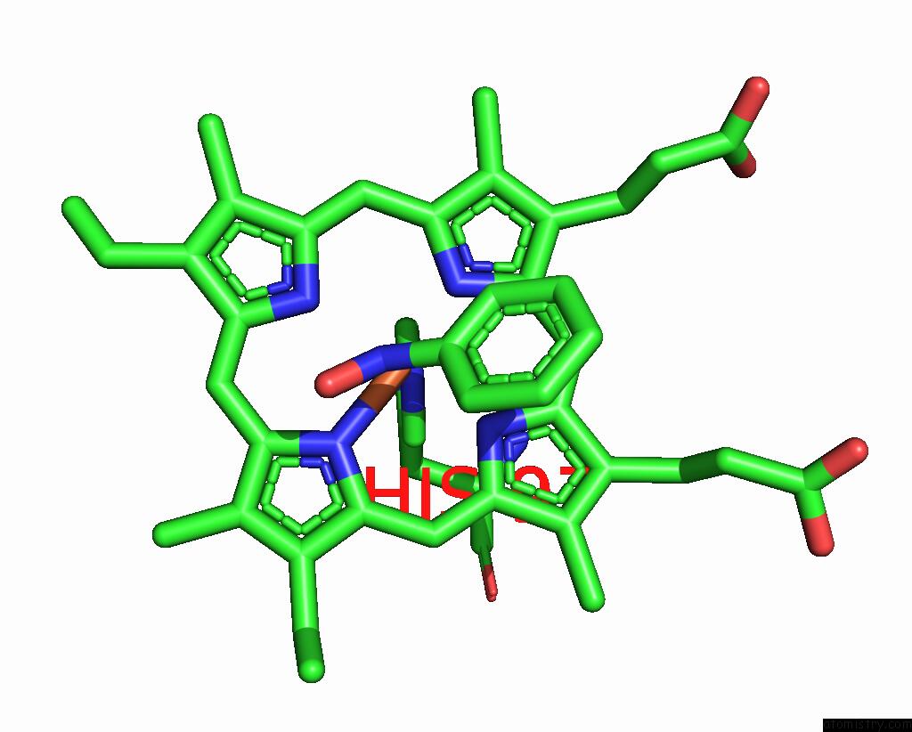
Mono view
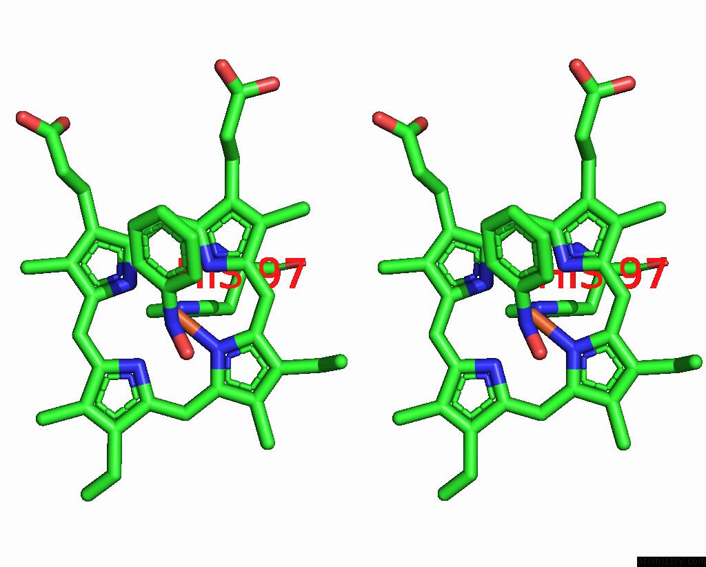
Stereo pair view
Mono view
Stereo pair view
A full contact list of Iron with other atoms in the Fe binding
site number 1 of X-Ray Structural Investigation of Leghemoglobin. VI. Structure of Acetate-Ferrileghemoglobin at A Resolution of 2.0 Angstroms (Russian) within 5.0Å range:
|
Reference:
E.G.Arutyunyan,
I.P.Kuranova,
B.K.Vainshtein,
W.Steigemann.
X-Ray Structural Investigation of Leghemoglobin. VI. Structure of Acetate-Ferrileghemoglobin at A Resolution of 2.0 Angstroms (Russian) Kristallografiya V. 25 80 1980.
ISSN: ISSN 0023-4761
Page generated: Sun Aug 4 00:24:13 2024
ISSN: ISSN 0023-4761
Last articles
F in 4RX5F in 4RV3
F in 4RV6
F in 4RUP
F in 4RRZ
F in 4RRW
F in 4RRX
F in 4RRS
F in 4RPL
F in 4RJD