Iron »
PDB 2l4d-2mta »
2mgd »
Iron in PDB 2mgd: High Resolution Crystal Structures of Five Distal Histidine Mutants of Sperm Whale Myoglobin
Protein crystallography data
The structure of High Resolution Crystal Structures of Five Distal Histidine Mutants of Sperm Whale Myoglobin, PDB code: 2mgd
was solved by
M.L.Quillin,
R.M.Arduini,
G.N.Phillips Jr.,
with X-Ray Crystallography technique. A brief refinement statistics is given in the table below:
| Resolution Low / High (Å) | 5.00 / 1.80 |
| Space group | P 6 |
| Cell size a, b, c (Å), α, β, γ (°) | 91.200, 91.200, 45.870, 90.00, 90.00, 120.00 |
| R / Rfree (%) | 15.9 / n/a |
Iron Binding Sites:
The binding sites of Iron atom in the High Resolution Crystal Structures of Five Distal Histidine Mutants of Sperm Whale Myoglobin
(pdb code 2mgd). This binding sites where shown within
5.0 Angstroms radius around Iron atom.
In total only one binding site of Iron was determined in the High Resolution Crystal Structures of Five Distal Histidine Mutants of Sperm Whale Myoglobin, PDB code: 2mgd:
In total only one binding site of Iron was determined in the High Resolution Crystal Structures of Five Distal Histidine Mutants of Sperm Whale Myoglobin, PDB code: 2mgd:
Iron binding site 1 out of 1 in 2mgd
Go back to
Iron binding site 1 out
of 1 in the High Resolution Crystal Structures of Five Distal Histidine Mutants of Sperm Whale Myoglobin
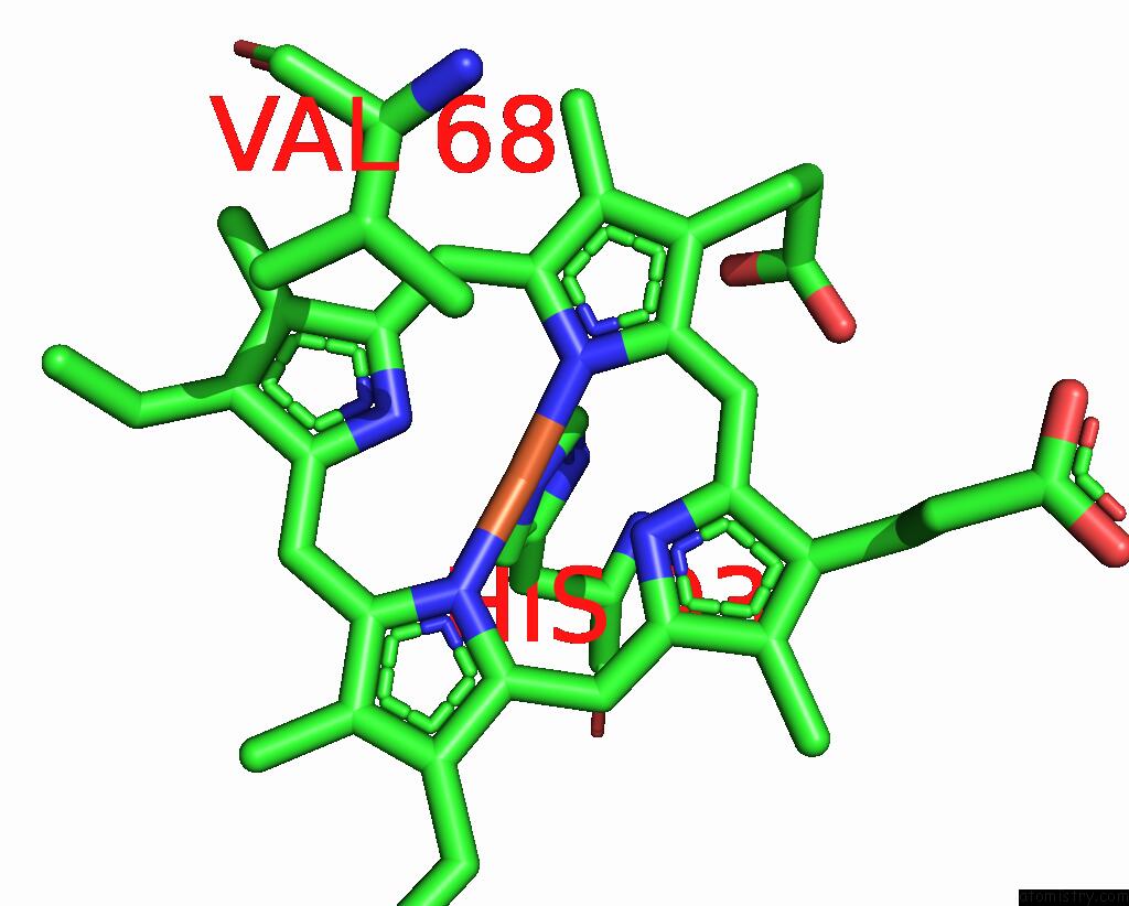
Mono view
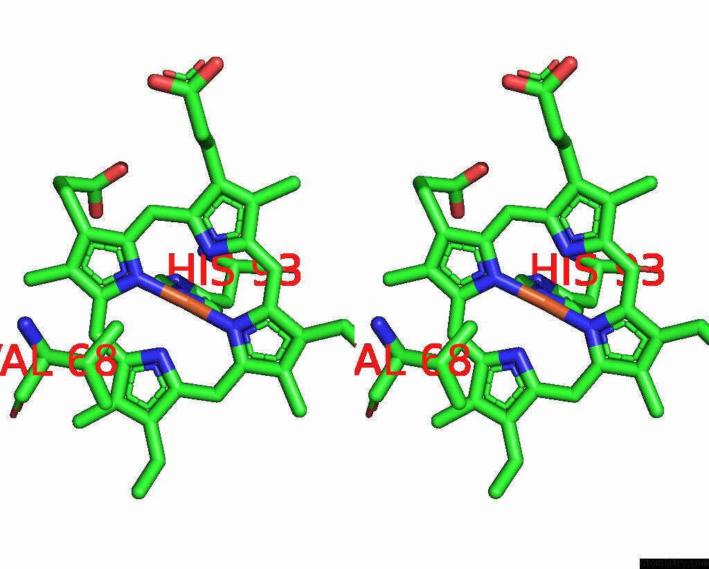
Stereo pair view
Mono view
Stereo pair view
A full contact list of Iron with other atoms in the Fe binding
site number 1 of High Resolution Crystal Structures of Five Distal Histidine Mutants of Sperm Whale Myoglobin within 5.0Å range:
|
Reference:
M.L.Quillin,
R.M.Arduini,
J.S.Olson,
G.N.Phillips Jr..
High-Resolution Crystal Structures of Distal Histidine Mutants of Sperm Whale Myoglobin. J.Mol.Biol. V. 234 140 1993.
ISSN: ISSN 0022-2836
PubMed: 8230194
DOI: 10.1006/JMBI.1993.1569
Page generated: Sun Aug 4 00:27:29 2024
ISSN: ISSN 0022-2836
PubMed: 8230194
DOI: 10.1006/JMBI.1993.1569
Last articles
Zn in 9MJ5Zn in 9HNW
Zn in 9G0L
Zn in 9FNE
Zn in 9DZN
Zn in 9E0I
Zn in 9D32
Zn in 9DAK
Zn in 8ZXC
Zn in 8ZUF