Iron »
PDB 2mya-2nwb »
2mye »
Iron in PDB 2mye: High Resolution X-Ray Structures of Myoglobin-and Hemoglobin-Alkyl Isocyanide Complexes
Protein crystallography data
The structure of High Resolution X-Ray Structures of Myoglobin-and Hemoglobin-Alkyl Isocyanide Complexes, PDB code: 2mye
was solved by
K.A.Johnson,
J.S.Olson,
G.N.Phillips Jr.,
with X-Ray Crystallography technique. A brief refinement statistics is given in the table below:
| Resolution Low / High (Å) | 5.00 / 1.68 |
| Space group | P 1 21 1 |
| Cell size a, b, c (Å), α, β, γ (°) | 64.640, 30.950, 34.950, 90.00, 106.08, 90.00 |
| R / Rfree (%) | 17.2 / n/a |
Iron Binding Sites:
The binding sites of Iron atom in the High Resolution X-Ray Structures of Myoglobin-and Hemoglobin-Alkyl Isocyanide Complexes
(pdb code 2mye). This binding sites where shown within
5.0 Angstroms radius around Iron atom.
In total only one binding site of Iron was determined in the High Resolution X-Ray Structures of Myoglobin-and Hemoglobin-Alkyl Isocyanide Complexes, PDB code: 2mye:
In total only one binding site of Iron was determined in the High Resolution X-Ray Structures of Myoglobin-and Hemoglobin-Alkyl Isocyanide Complexes, PDB code: 2mye:
Iron binding site 1 out of 1 in 2mye
Go back to
Iron binding site 1 out
of 1 in the High Resolution X-Ray Structures of Myoglobin-and Hemoglobin-Alkyl Isocyanide Complexes
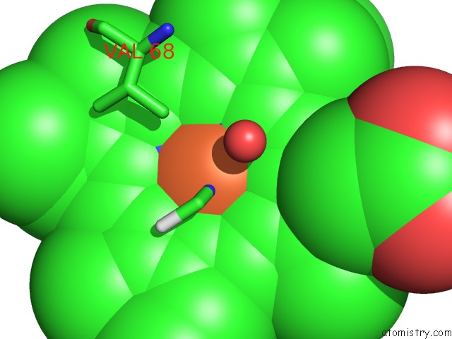
Mono view
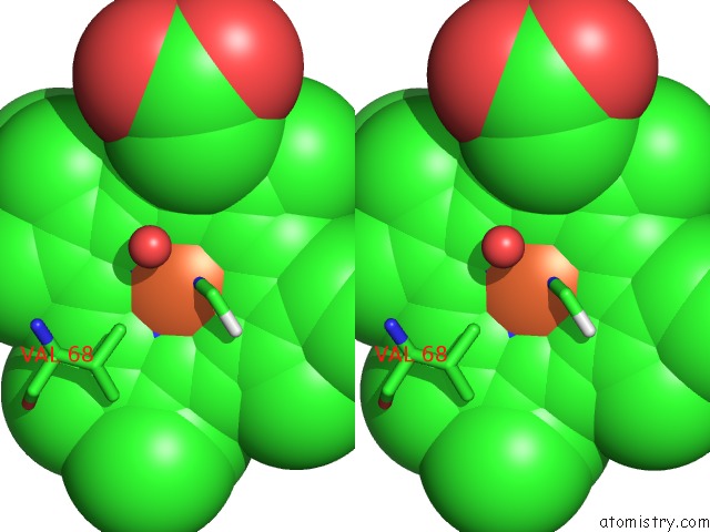
Stereo pair view
Mono view
Stereo pair view
A full contact list of Iron with other atoms in the Fe binding
site number 1 of High Resolution X-Ray Structures of Myoglobin-and Hemoglobin-Alkyl Isocyanide Complexes within 5.0Å range:
|
Reference:
K.A.Johnson,
K.A.Johnson,
J.S.Olson,
G.N.Phillips Jr..
N/A N/A.
Page generated: Thu Jul 17 02:59:09 2025
Last articles
Fe in 2YXOFe in 2YRS
Fe in 2YXC
Fe in 2YNM
Fe in 2YVJ
Fe in 2YP1
Fe in 2YU2
Fe in 2YU1
Fe in 2YQB
Fe in 2YOO