Iron »
PDB 2mya-2nwb »
2nox »
Iron in PDB 2nox: Crystal Structure of Tryptophan 2,3-Dioxygenase From Ralstonia Metallidurans
Enzymatic activity of Crystal Structure of Tryptophan 2,3-Dioxygenase From Ralstonia Metallidurans
All present enzymatic activity of Crystal Structure of Tryptophan 2,3-Dioxygenase From Ralstonia Metallidurans:
1.13.11.11;
1.13.11.11;
Protein crystallography data
The structure of Crystal Structure of Tryptophan 2,3-Dioxygenase From Ralstonia Metallidurans, PDB code: 2nox
was solved by
Y.Zhang,
S.A.Kang,
T.Mukherjee,
S.Bale,
B.R.Crane,
T.P.Begley,
S.E.Ealick,
with X-Ray Crystallography technique. A brief refinement statistics is given in the table below:
| Resolution Low / High (Å) | 50.12 / 2.40 |
| Space group | P 1 |
| Cell size a, b, c (Å), α, β, γ (°) | 72.544, 132.121, 139.950, 66.97, 85.06, 89.89 |
| R / Rfree (%) | 21 / 27 |
Iron Binding Sites:
Pages:
>>> Page 1 <<< Page 2, Binding sites: 11 - 16;Binding sites:
The binding sites of Iron atom in the Crystal Structure of Tryptophan 2,3-Dioxygenase From Ralstonia Metallidurans (pdb code 2nox). This binding sites where shown within 5.0 Angstroms radius around Iron atom.In total 16 binding sites of Iron where determined in the Crystal Structure of Tryptophan 2,3-Dioxygenase From Ralstonia Metallidurans, PDB code: 2nox:
Jump to Iron binding site number: 1; 2; 3; 4; 5; 6; 7; 8; 9; 10;
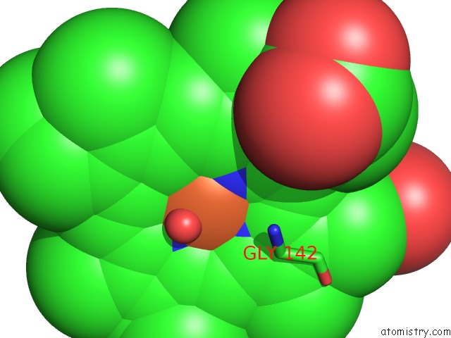
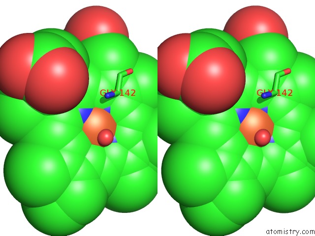
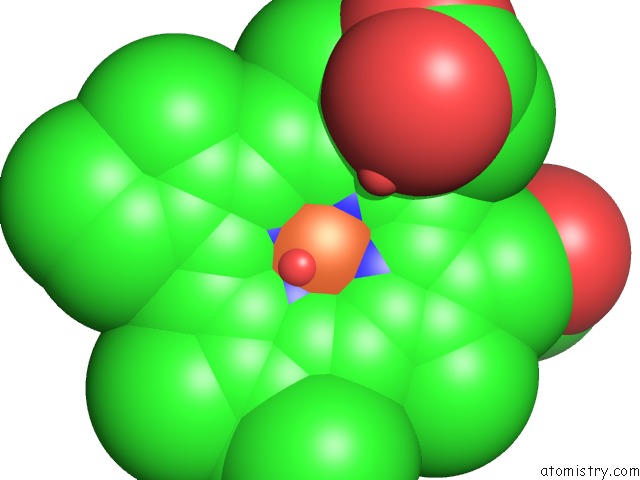
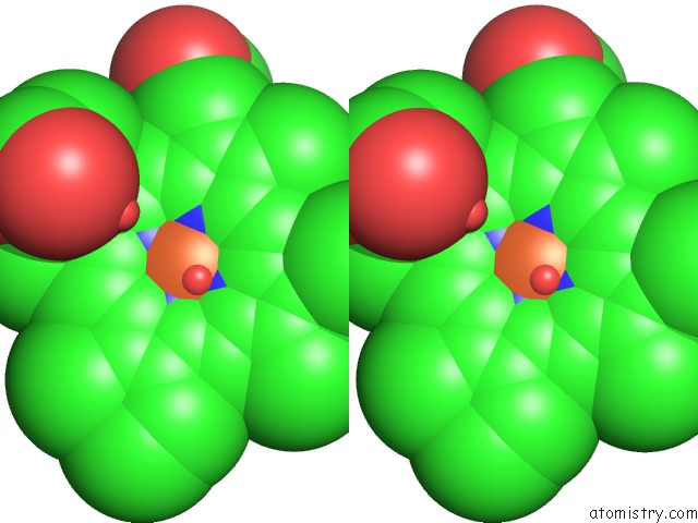
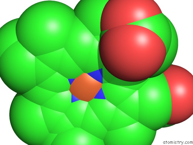
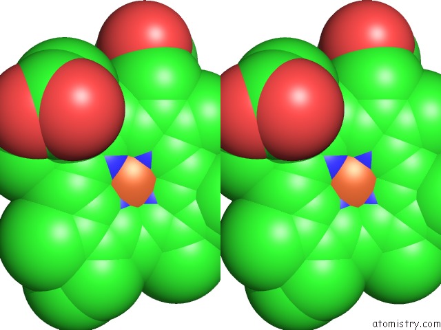
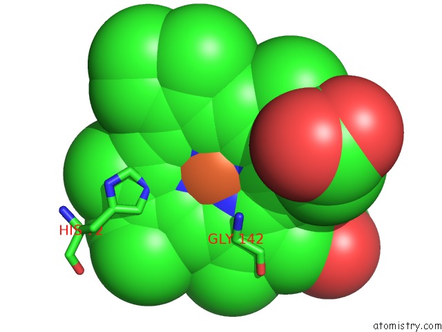
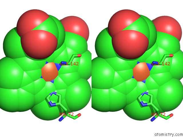
Iron binding site 1 out of 16 in 2nox
Go back to
Iron binding site 1 out
of 16 in the Crystal Structure of Tryptophan 2,3-Dioxygenase From Ralstonia Metallidurans
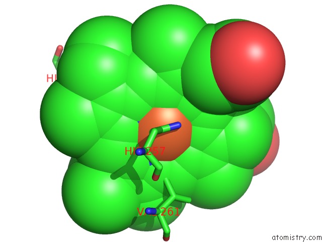
Mono view
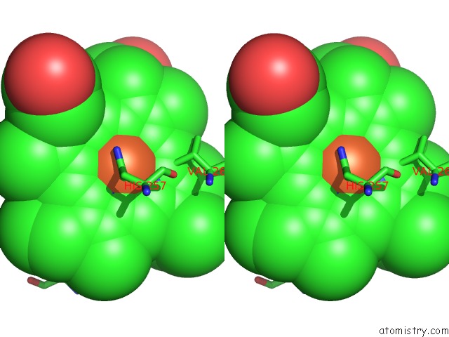
Stereo pair view
Mono view
Stereo pair view
A full contact list of Iron with other atoms in the Fe binding
site number 1 of Crystal Structure of Tryptophan 2,3-Dioxygenase From Ralstonia Metallidurans within 5.0Å range:
|
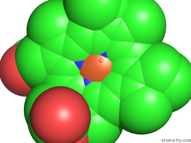
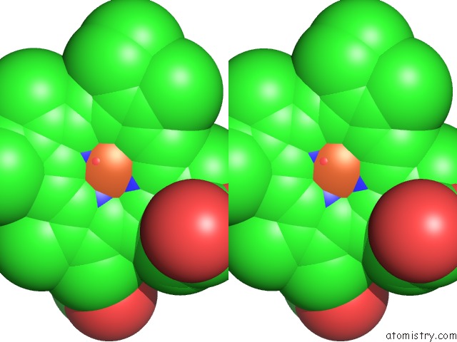
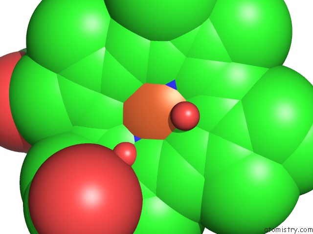
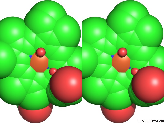
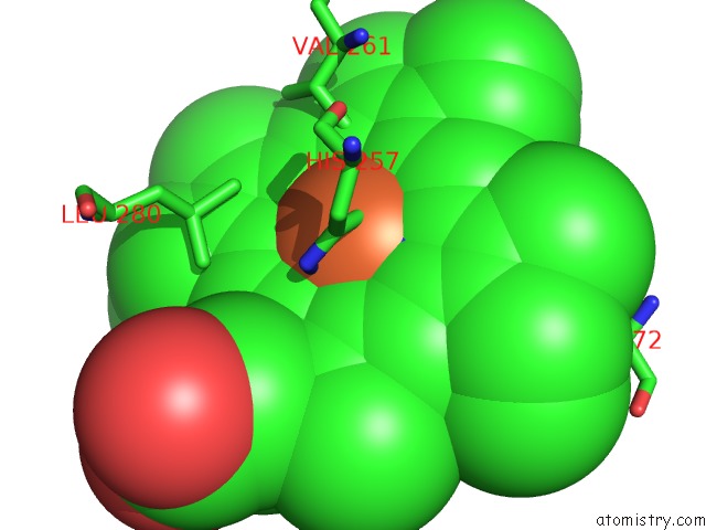
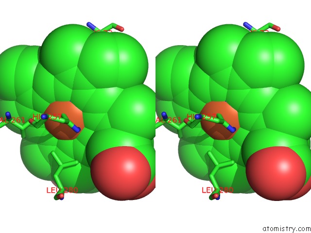
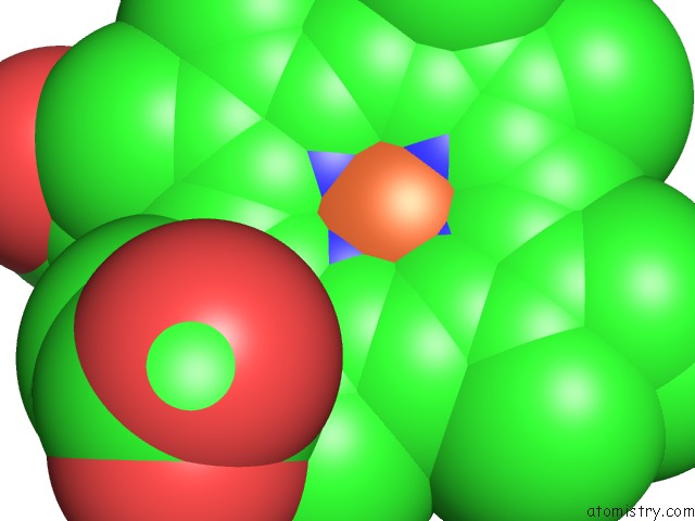
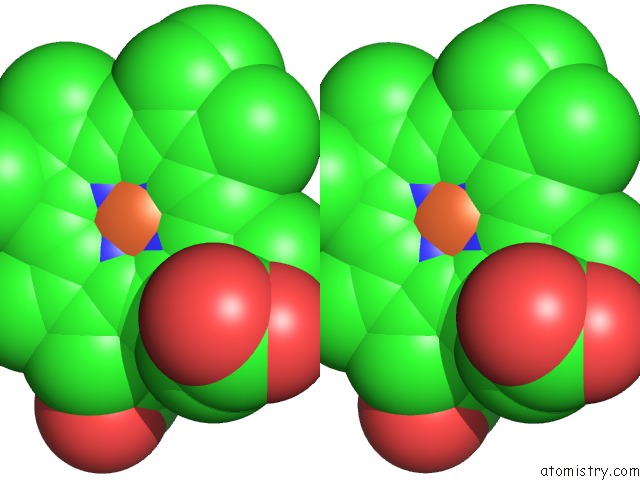
Iron binding site 2 out of 16 in 2nox
Go back to
Iron binding site 2 out
of 16 in the Crystal Structure of Tryptophan 2,3-Dioxygenase From Ralstonia Metallidurans
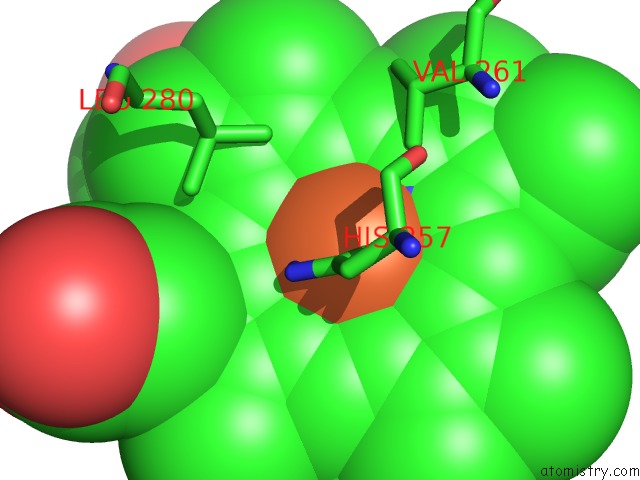
Mono view
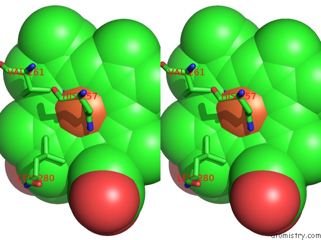
Stereo pair view
Mono view
Stereo pair view
A full contact list of Iron with other atoms in the Fe binding
site number 2 of Crystal Structure of Tryptophan 2,3-Dioxygenase From Ralstonia Metallidurans within 5.0Å range:
|
Iron binding site 3 out of 16 in 2nox
Go back to
Iron binding site 3 out
of 16 in the Crystal Structure of Tryptophan 2,3-Dioxygenase From Ralstonia Metallidurans
Mono view
Stereo pair view
Mono view
Stereo pair view
A full contact list of Iron with other atoms in the Fe binding
site number 3 of Crystal Structure of Tryptophan 2,3-Dioxygenase From Ralstonia Metallidurans within 5.0Å range:
|
Iron binding site 4 out of 16 in 2nox
Go back to
Iron binding site 4 out
of 16 in the Crystal Structure of Tryptophan 2,3-Dioxygenase From Ralstonia Metallidurans
Mono view
Stereo pair view
Mono view
Stereo pair view
A full contact list of Iron with other atoms in the Fe binding
site number 4 of Crystal Structure of Tryptophan 2,3-Dioxygenase From Ralstonia Metallidurans within 5.0Å range:
|
Iron binding site 5 out of 16 in 2nox
Go back to
Iron binding site 5 out
of 16 in the Crystal Structure of Tryptophan 2,3-Dioxygenase From Ralstonia Metallidurans
Mono view
Stereo pair view
Mono view
Stereo pair view
A full contact list of Iron with other atoms in the Fe binding
site number 5 of Crystal Structure of Tryptophan 2,3-Dioxygenase From Ralstonia Metallidurans within 5.0Å range:
|
Iron binding site 6 out of 16 in 2nox
Go back to
Iron binding site 6 out
of 16 in the Crystal Structure of Tryptophan 2,3-Dioxygenase From Ralstonia Metallidurans
Mono view
Stereo pair view
Mono view
Stereo pair view
A full contact list of Iron with other atoms in the Fe binding
site number 6 of Crystal Structure of Tryptophan 2,3-Dioxygenase From Ralstonia Metallidurans within 5.0Å range:
|
Iron binding site 7 out of 16 in 2nox
Go back to
Iron binding site 7 out
of 16 in the Crystal Structure of Tryptophan 2,3-Dioxygenase From Ralstonia Metallidurans
Mono view
Stereo pair view
Mono view
Stereo pair view
A full contact list of Iron with other atoms in the Fe binding
site number 7 of Crystal Structure of Tryptophan 2,3-Dioxygenase From Ralstonia Metallidurans within 5.0Å range:
|
Iron binding site 8 out of 16 in 2nox
Go back to
Iron binding site 8 out
of 16 in the Crystal Structure of Tryptophan 2,3-Dioxygenase From Ralstonia Metallidurans
Mono view
Stereo pair view
Mono view
Stereo pair view
A full contact list of Iron with other atoms in the Fe binding
site number 8 of Crystal Structure of Tryptophan 2,3-Dioxygenase From Ralstonia Metallidurans within 5.0Å range:
|
Iron binding site 9 out of 16 in 2nox
Go back to
Iron binding site 9 out
of 16 in the Crystal Structure of Tryptophan 2,3-Dioxygenase From Ralstonia Metallidurans
Mono view
Stereo pair view
Mono view
Stereo pair view
A full contact list of Iron with other atoms in the Fe binding
site number 9 of Crystal Structure of Tryptophan 2,3-Dioxygenase From Ralstonia Metallidurans within 5.0Å range:
|
Iron binding site 10 out of 16 in 2nox
Go back to
Iron binding site 10 out
of 16 in the Crystal Structure of Tryptophan 2,3-Dioxygenase From Ralstonia Metallidurans
Mono view
Stereo pair view
Mono view
Stereo pair view
A full contact list of Iron with other atoms in the Fe binding
site number 10 of Crystal Structure of Tryptophan 2,3-Dioxygenase From Ralstonia Metallidurans within 5.0Å range:
|
Reference:
Y.Zhang,
S.A.Kang,
T.Mukherjee,
S.Bale,
B.R.Crane,
T.P.Begley,
S.E.Ealick.
Crystal Structure and Mechanism of Tryptophan 2,3-Dioxygenase, A Heme Enzyme Involved in Tryptophan Catabolism and in Quinolinate Biosynthesis. Biochemistry V. 46 145 2007.
ISSN: ISSN 0006-2960
PubMed: 17198384
DOI: 10.1021/BI0620095
Page generated: Thu Jul 17 03:02:33 2025
ISSN: ISSN 0006-2960
PubMed: 17198384
DOI: 10.1021/BI0620095
Last articles
Fe in 2YXOFe in 2YRS
Fe in 2YXC
Fe in 2YNM
Fe in 2YVJ
Fe in 2YP1
Fe in 2YU2
Fe in 2YU1
Fe in 2YQB
Fe in 2YOO