Iron »
PDB 2nwf-2oof »
2nya »
Iron in PDB 2nya: Crystal Structure of the Periplasmic Nitrate Reductase (Nap) From Escherichia Coli
Enzymatic activity of Crystal Structure of the Periplasmic Nitrate Reductase (Nap) From Escherichia Coli
All present enzymatic activity of Crystal Structure of the Periplasmic Nitrate Reductase (Nap) From Escherichia Coli:
1.7.99.4;
1.7.99.4;
Protein crystallography data
The structure of Crystal Structure of the Periplasmic Nitrate Reductase (Nap) From Escherichia Coli, PDB code: 2nya
was solved by
B.J.N.Jepson,
D.J.Richardson,
A.M.Hemmings,
with X-Ray Crystallography technique. A brief refinement statistics is given in the table below:
| Resolution Low / High (Å) | 50.00 / 2.50 |
| Space group | P 1 21 1 |
| Cell size a, b, c (Å), α, β, γ (°) | 69.449, 94.596, 131.211, 90.00, 96.34, 90.00 |
| R / Rfree (%) | 18.1 / 24.3 |
Other elements in 2nya:
The structure of Crystal Structure of the Periplasmic Nitrate Reductase (Nap) From Escherichia Coli also contains other interesting chemical elements:
| Molybdenum | (Mo) | 2 atoms |
Iron Binding Sites:
The binding sites of Iron atom in the Crystal Structure of the Periplasmic Nitrate Reductase (Nap) From Escherichia Coli
(pdb code 2nya). This binding sites where shown within
5.0 Angstroms radius around Iron atom.
In total 8 binding sites of Iron where determined in the Crystal Structure of the Periplasmic Nitrate Reductase (Nap) From Escherichia Coli, PDB code: 2nya:
Jump to Iron binding site number: 1; 2; 3; 4; 5; 6; 7; 8;
In total 8 binding sites of Iron where determined in the Crystal Structure of the Periplasmic Nitrate Reductase (Nap) From Escherichia Coli, PDB code: 2nya:
Jump to Iron binding site number: 1; 2; 3; 4; 5; 6; 7; 8;
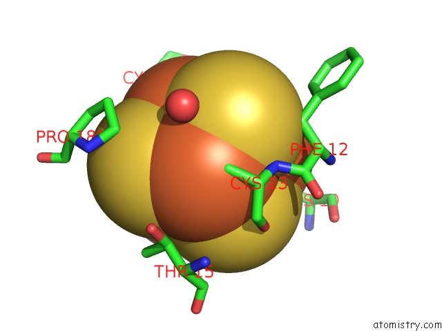
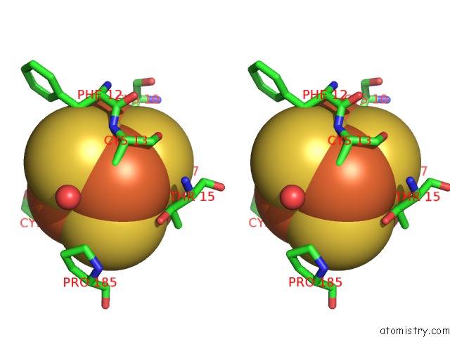
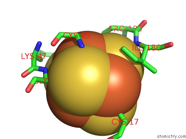
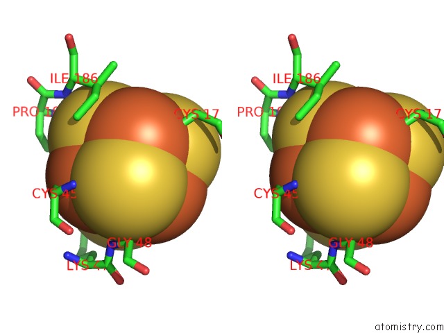
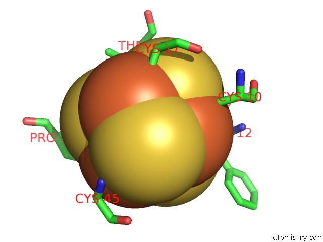
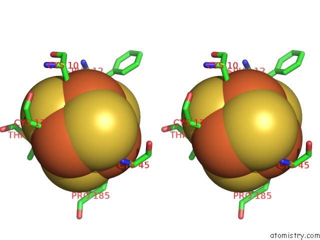
Iron binding site 1 out of 8 in 2nya
Go back to
Iron binding site 1 out
of 8 in the Crystal Structure of the Periplasmic Nitrate Reductase (Nap) From Escherichia Coli
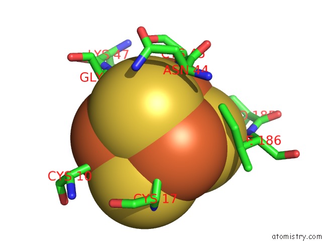
Mono view
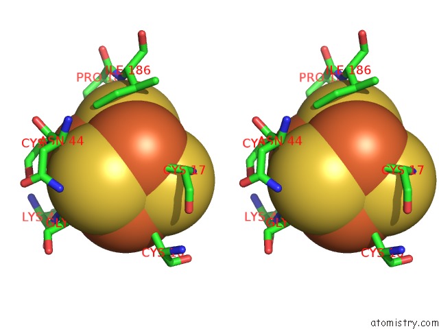
Stereo pair view
Mono view
Stereo pair view
A full contact list of Iron with other atoms in the Fe binding
site number 1 of Crystal Structure of the Periplasmic Nitrate Reductase (Nap) From Escherichia Coli within 5.0Å range:
|
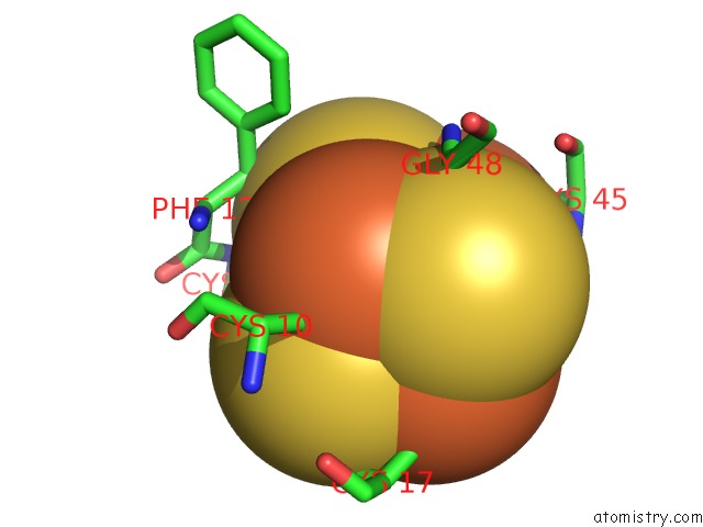
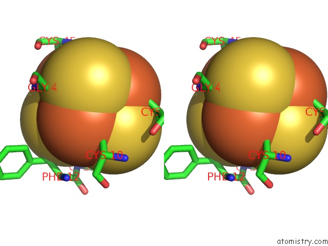
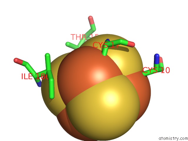
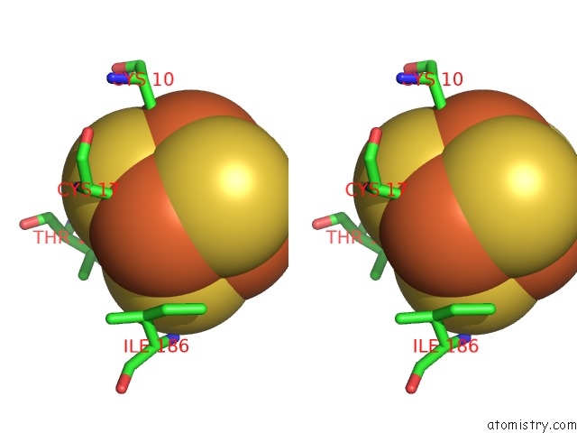
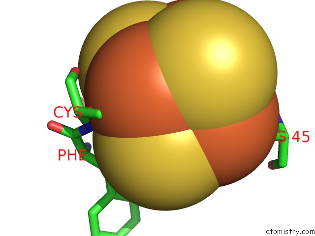
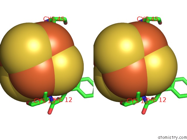
Iron binding site 2 out of 8 in 2nya
Go back to
Iron binding site 2 out
of 8 in the Crystal Structure of the Periplasmic Nitrate Reductase (Nap) From Escherichia Coli
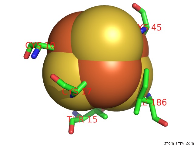
Mono view
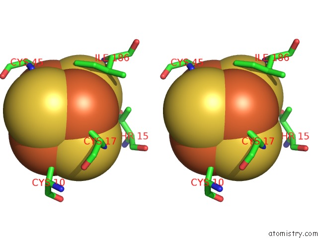
Stereo pair view
Mono view
Stereo pair view
A full contact list of Iron with other atoms in the Fe binding
site number 2 of Crystal Structure of the Periplasmic Nitrate Reductase (Nap) From Escherichia Coli within 5.0Å range:
|
Iron binding site 3 out of 8 in 2nya
Go back to
Iron binding site 3 out
of 8 in the Crystal Structure of the Periplasmic Nitrate Reductase (Nap) From Escherichia Coli
Mono view
Stereo pair view
Mono view
Stereo pair view
A full contact list of Iron with other atoms in the Fe binding
site number 3 of Crystal Structure of the Periplasmic Nitrate Reductase (Nap) From Escherichia Coli within 5.0Å range:
|
Iron binding site 4 out of 8 in 2nya
Go back to
Iron binding site 4 out
of 8 in the Crystal Structure of the Periplasmic Nitrate Reductase (Nap) From Escherichia Coli
Mono view
Stereo pair view
Mono view
Stereo pair view
A full contact list of Iron with other atoms in the Fe binding
site number 4 of Crystal Structure of the Periplasmic Nitrate Reductase (Nap) From Escherichia Coli within 5.0Å range:
|
Iron binding site 5 out of 8 in 2nya
Go back to
Iron binding site 5 out
of 8 in the Crystal Structure of the Periplasmic Nitrate Reductase (Nap) From Escherichia Coli
Mono view
Stereo pair view
Mono view
Stereo pair view
A full contact list of Iron with other atoms in the Fe binding
site number 5 of Crystal Structure of the Periplasmic Nitrate Reductase (Nap) From Escherichia Coli within 5.0Å range:
|
Iron binding site 6 out of 8 in 2nya
Go back to
Iron binding site 6 out
of 8 in the Crystal Structure of the Periplasmic Nitrate Reductase (Nap) From Escherichia Coli
Mono view
Stereo pair view
Mono view
Stereo pair view
A full contact list of Iron with other atoms in the Fe binding
site number 6 of Crystal Structure of the Periplasmic Nitrate Reductase (Nap) From Escherichia Coli within 5.0Å range:
|
Iron binding site 7 out of 8 in 2nya
Go back to
Iron binding site 7 out
of 8 in the Crystal Structure of the Periplasmic Nitrate Reductase (Nap) From Escherichia Coli
Mono view
Stereo pair view
Mono view
Stereo pair view
A full contact list of Iron with other atoms in the Fe binding
site number 7 of Crystal Structure of the Periplasmic Nitrate Reductase (Nap) From Escherichia Coli within 5.0Å range:
|
Iron binding site 8 out of 8 in 2nya
Go back to
Iron binding site 8 out
of 8 in the Crystal Structure of the Periplasmic Nitrate Reductase (Nap) From Escherichia Coli
Mono view
Stereo pair view
Mono view
Stereo pair view
A full contact list of Iron with other atoms in the Fe binding
site number 8 of Crystal Structure of the Periplasmic Nitrate Reductase (Nap) From Escherichia Coli within 5.0Å range:
|
Reference:
B.J.Jepson,
S.Mohan,
T.A.Clarke,
A.J.Gates,
J.A.Cole,
C.S.Butler,
J.N.Butt,
A.M.Hemmings,
D.J.Richardson.
Spectropotentiometric and Structural Analysis of the Periplasmic Nitrate Reductase From Escherichia Coli J.Biol.Chem. V. 282 6425 2007.
ISSN: ISSN 0021-9258
PubMed: 17130127
DOI: 10.1074/JBC.M607353200
Page generated: Thu Jul 17 03:06:40 2025
ISSN: ISSN 0021-9258
PubMed: 17130127
DOI: 10.1074/JBC.M607353200
Last articles
Fe in 2YXOFe in 2YRS
Fe in 2YXC
Fe in 2YNM
Fe in 2YVJ
Fe in 2YP1
Fe in 2YU2
Fe in 2YU1
Fe in 2YQB
Fe in 2YOO