Iron »
PDB 2nwf-2oof »
2o7u »
Iron in PDB 2o7u: Crystal Structure of K206E/K296E Mutant of the N-Terminal Half Molecule of Human Transferrin
Protein crystallography data
The structure of Crystal Structure of K206E/K296E Mutant of the N-Terminal Half Molecule of Human Transferrin, PDB code: 2o7u
was solved by
H.M.Baker,
D.Nurizzo,
A.B.Mason,
E.N.Baker,
with X-Ray Crystallography technique. A brief refinement statistics is given in the table below:
| Resolution Low / High (Å) | 29.76 / 2.80 |
| Space group | C 1 2 1 |
| Cell size a, b, c (Å), α, β, γ (°) | 169.462, 97.897, 208.953, 90.00, 90.01, 90.00 |
| R / Rfree (%) | 23 / 25.9 |
Iron Binding Sites:
The binding sites of Iron atom in the Crystal Structure of K206E/K296E Mutant of the N-Terminal Half Molecule of Human Transferrin
(pdb code 2o7u). This binding sites where shown within
5.0 Angstroms radius around Iron atom.
In total 9 binding sites of Iron where determined in the Crystal Structure of K206E/K296E Mutant of the N-Terminal Half Molecule of Human Transferrin, PDB code: 2o7u:
Jump to Iron binding site number: 1; 2; 3; 4; 5; 6; 7; 8; 9;
In total 9 binding sites of Iron where determined in the Crystal Structure of K206E/K296E Mutant of the N-Terminal Half Molecule of Human Transferrin, PDB code: 2o7u:
Jump to Iron binding site number: 1; 2; 3; 4; 5; 6; 7; 8; 9;
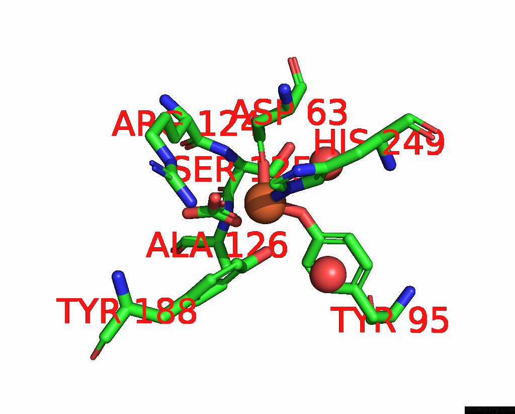
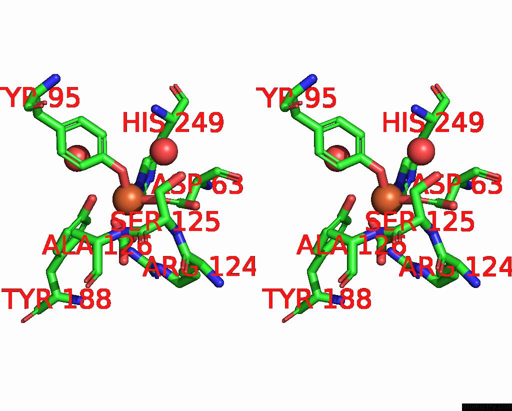
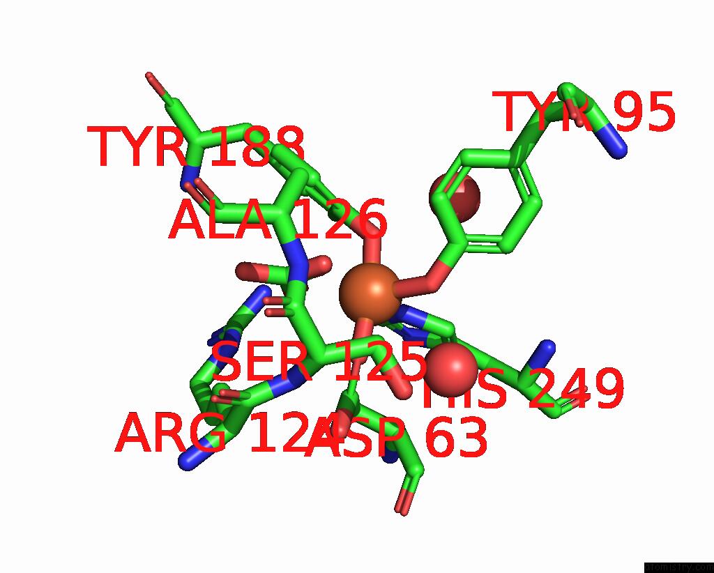
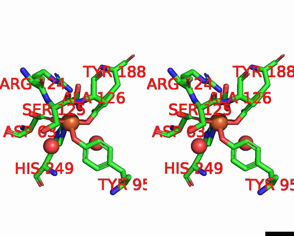
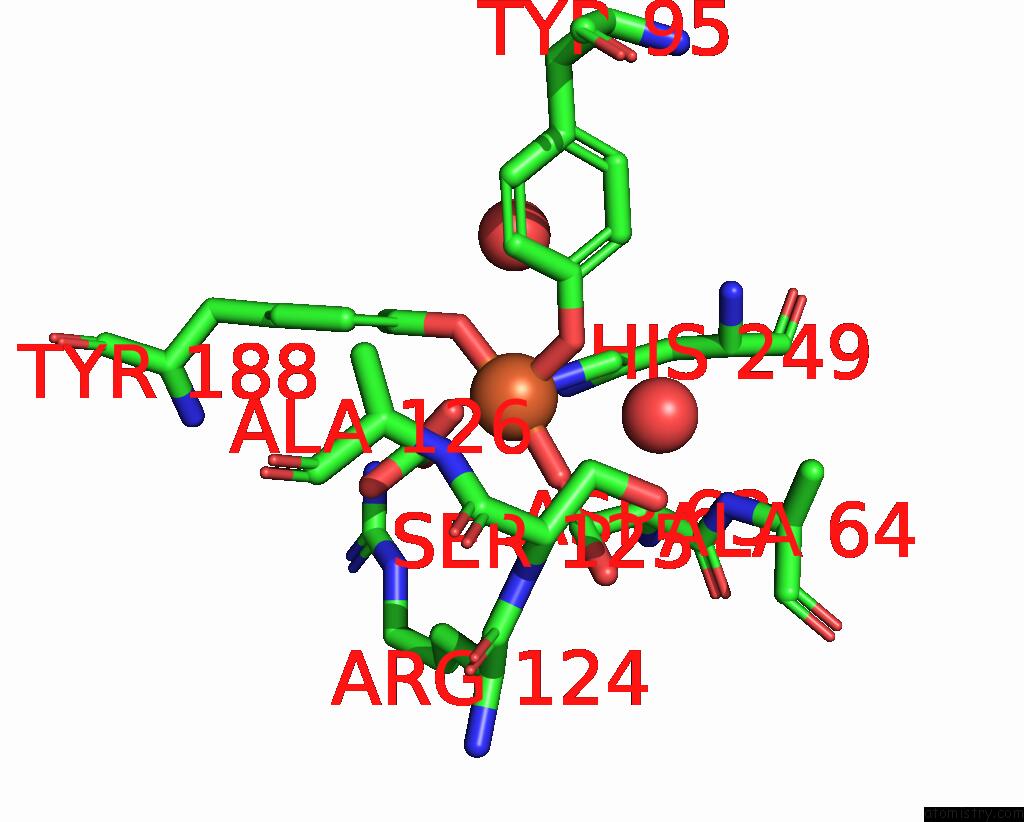
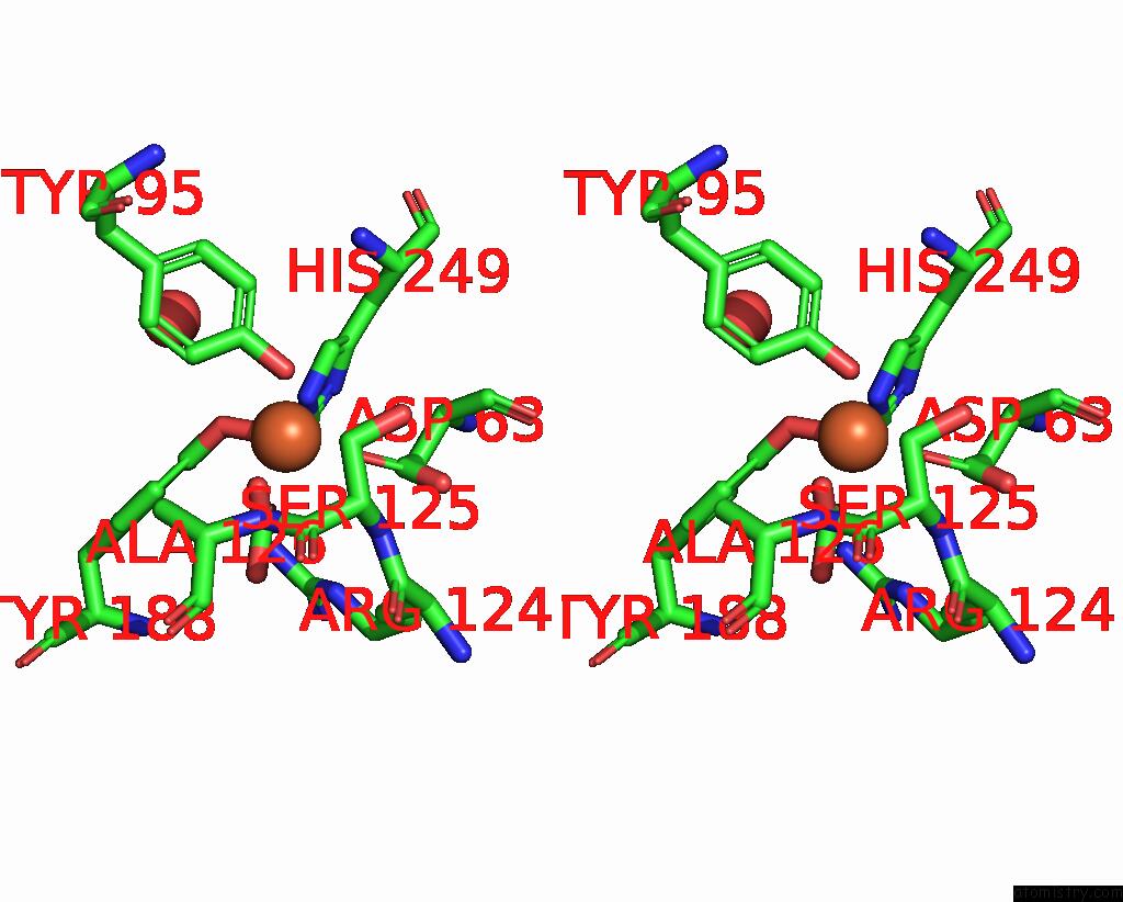
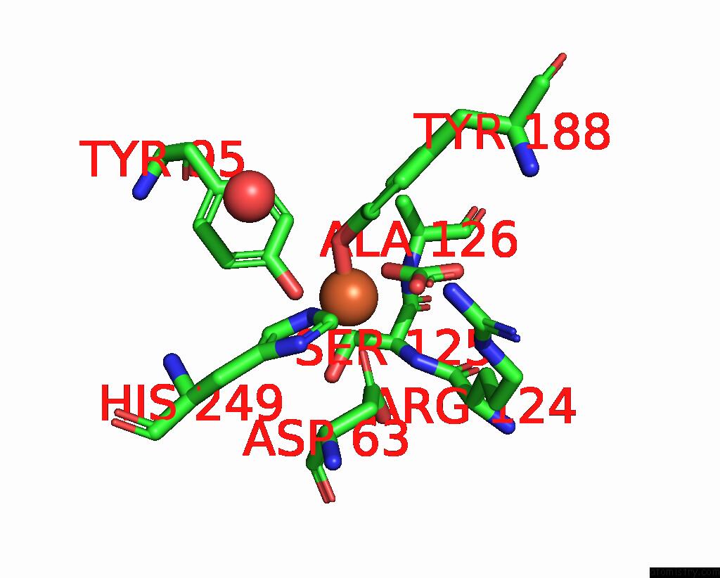
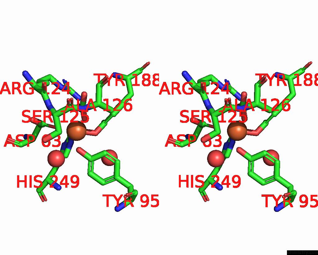
Iron binding site 1 out of 9 in 2o7u
Go back to
Iron binding site 1 out
of 9 in the Crystal Structure of K206E/K296E Mutant of the N-Terminal Half Molecule of Human Transferrin

Mono view
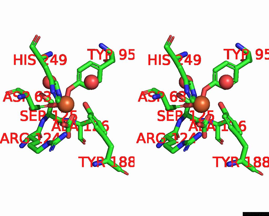
Stereo pair view
Mono view
Stereo pair view
A full contact list of Iron with other atoms in the Fe binding
site number 1 of Crystal Structure of K206E/K296E Mutant of the N-Terminal Half Molecule of Human Transferrin within 5.0Å range:
|
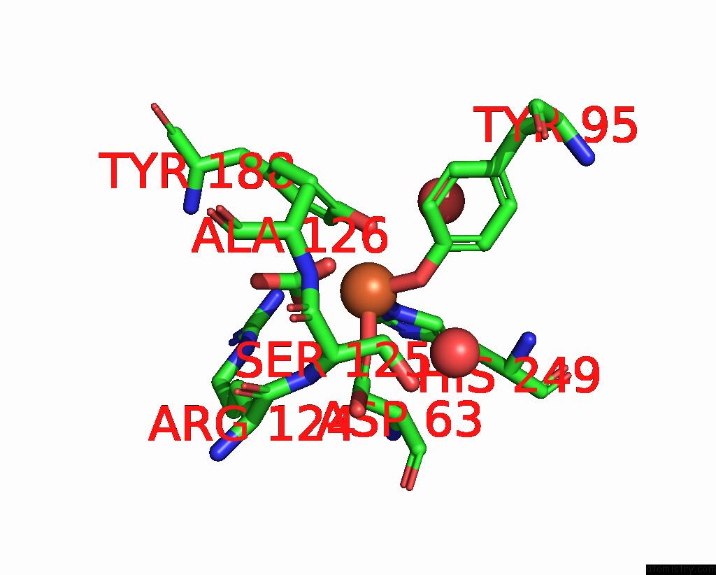
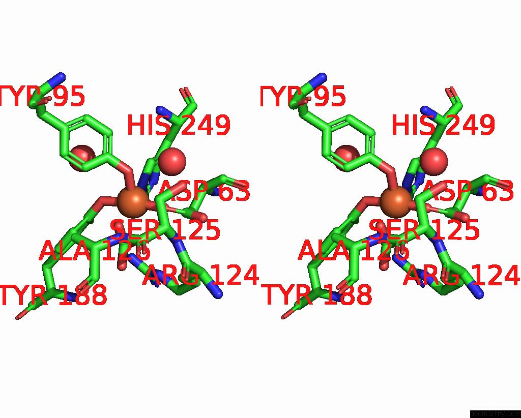
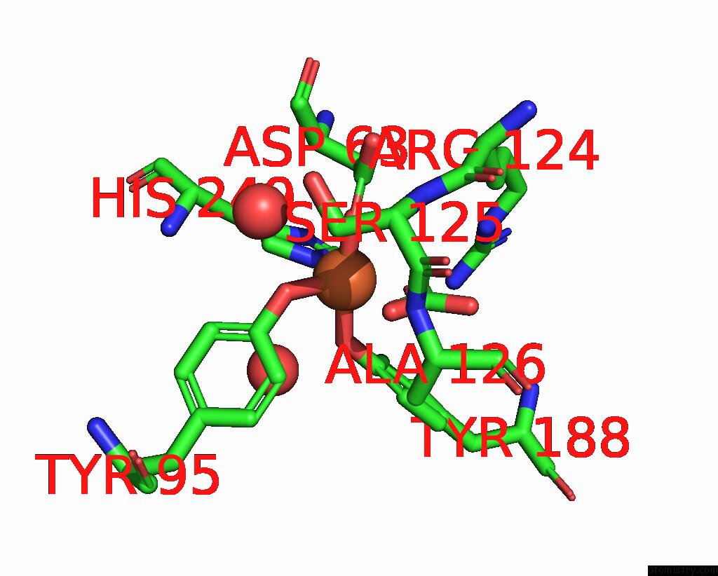
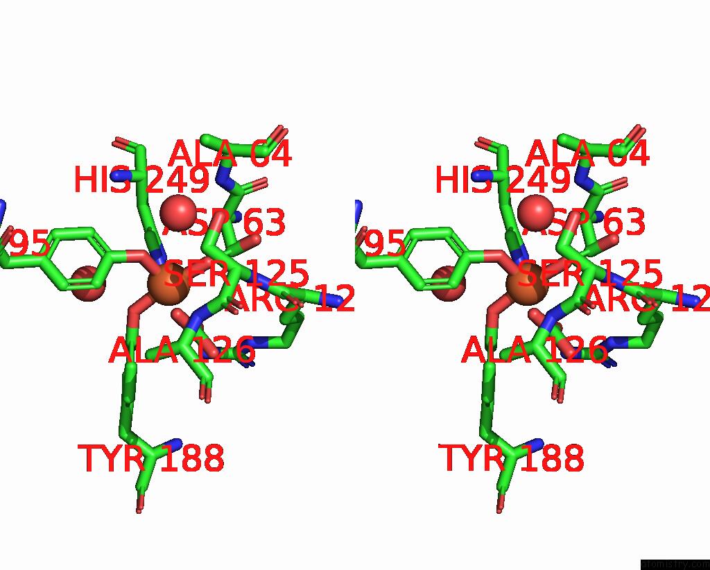
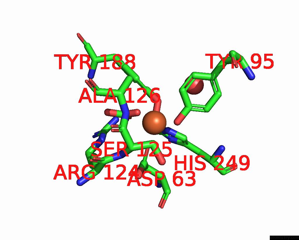
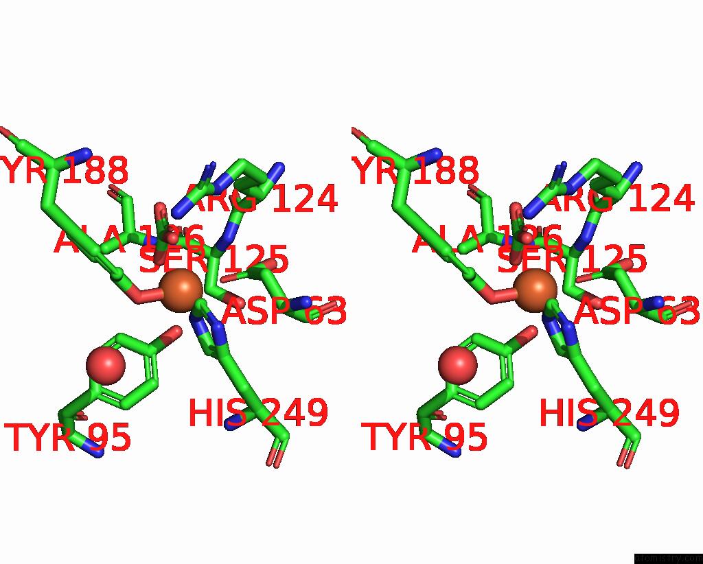
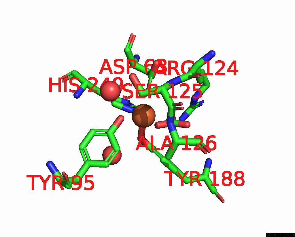
Iron binding site 2 out of 9 in 2o7u
Go back to
Iron binding site 2 out
of 9 in the Crystal Structure of K206E/K296E Mutant of the N-Terminal Half Molecule of Human Transferrin
Mono view
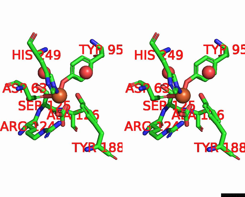
Stereo pair view
Mono view
Stereo pair view
A full contact list of Iron with other atoms in the Fe binding
site number 2 of Crystal Structure of K206E/K296E Mutant of the N-Terminal Half Molecule of Human Transferrin within 5.0Å range:
|
Iron binding site 3 out of 9 in 2o7u
Go back to
Iron binding site 3 out
of 9 in the Crystal Structure of K206E/K296E Mutant of the N-Terminal Half Molecule of Human Transferrin
Mono view
Stereo pair view
Mono view
Stereo pair view
A full contact list of Iron with other atoms in the Fe binding
site number 3 of Crystal Structure of K206E/K296E Mutant of the N-Terminal Half Molecule of Human Transferrin within 5.0Å range:
|
Iron binding site 4 out of 9 in 2o7u
Go back to
Iron binding site 4 out
of 9 in the Crystal Structure of K206E/K296E Mutant of the N-Terminal Half Molecule of Human Transferrin
Mono view
Stereo pair view
Mono view
Stereo pair view
A full contact list of Iron with other atoms in the Fe binding
site number 4 of Crystal Structure of K206E/K296E Mutant of the N-Terminal Half Molecule of Human Transferrin within 5.0Å range:
|
Iron binding site 5 out of 9 in 2o7u
Go back to
Iron binding site 5 out
of 9 in the Crystal Structure of K206E/K296E Mutant of the N-Terminal Half Molecule of Human Transferrin
Mono view
Stereo pair view
Mono view
Stereo pair view
A full contact list of Iron with other atoms in the Fe binding
site number 5 of Crystal Structure of K206E/K296E Mutant of the N-Terminal Half Molecule of Human Transferrin within 5.0Å range:
|
Iron binding site 6 out of 9 in 2o7u
Go back to
Iron binding site 6 out
of 9 in the Crystal Structure of K206E/K296E Mutant of the N-Terminal Half Molecule of Human Transferrin
Mono view
Stereo pair view
Mono view
Stereo pair view
A full contact list of Iron with other atoms in the Fe binding
site number 6 of Crystal Structure of K206E/K296E Mutant of the N-Terminal Half Molecule of Human Transferrin within 5.0Å range:
|
Iron binding site 7 out of 9 in 2o7u
Go back to
Iron binding site 7 out
of 9 in the Crystal Structure of K206E/K296E Mutant of the N-Terminal Half Molecule of Human Transferrin
Mono view
Stereo pair view
Mono view
Stereo pair view
A full contact list of Iron with other atoms in the Fe binding
site number 7 of Crystal Structure of K206E/K296E Mutant of the N-Terminal Half Molecule of Human Transferrin within 5.0Å range:
|
Iron binding site 8 out of 9 in 2o7u
Go back to
Iron binding site 8 out
of 9 in the Crystal Structure of K206E/K296E Mutant of the N-Terminal Half Molecule of Human Transferrin
Mono view
Stereo pair view
Mono view
Stereo pair view
A full contact list of Iron with other atoms in the Fe binding
site number 8 of Crystal Structure of K206E/K296E Mutant of the N-Terminal Half Molecule of Human Transferrin within 5.0Å range:
|
Iron binding site 9 out of 9 in 2o7u
Go back to
Iron binding site 9 out
of 9 in the Crystal Structure of K206E/K296E Mutant of the N-Terminal Half Molecule of Human Transferrin
Mono view
Stereo pair view
Mono view
Stereo pair view
A full contact list of Iron with other atoms in the Fe binding
site number 9 of Crystal Structure of K206E/K296E Mutant of the N-Terminal Half Molecule of Human Transferrin within 5.0Å range:
|
Reference:
H.M.Baker,
D.Nurizzo,
A.B.Mason,
E.N.Baker.
Structures of Two Mutants That Probe the Role in Iron Release of the Dilysine Pair in the N-Lobe of Human Transferrin. Acta Crystallogr.,Sect.D V. 63 408 2007.
ISSN: ISSN 0907-4449
PubMed: 17327678
DOI: 10.1107/S0907444907000182
Page generated: Sun Aug 4 00:56:46 2024
ISSN: ISSN 0907-4449
PubMed: 17327678
DOI: 10.1107/S0907444907000182
Last articles
Zn in 9MJ5Zn in 9HNW
Zn in 9G0L
Zn in 9FNE
Zn in 9DZN
Zn in 9E0I
Zn in 9D32
Zn in 9DAK
Zn in 8ZXC
Zn in 8ZUF