Iron »
PDB 2nwf-2oof »
2oh9 »
Iron in PDB 2oh9: Myoglobin Cavity Mutant V68W
Protein crystallography data
The structure of Myoglobin Cavity Mutant V68W, PDB code: 2oh9
was solved by
G.N.Phillips Jr.,
T.Li,
E.A.Brucker,
J.Soman,
J.S.Olson,
with X-Ray Crystallography technique. A brief refinement statistics is given in the table below:
| Resolution Low / High (Å) | 45.89 / 1.80 |
| Space group | P 6 |
| Cell size a, b, c (Å), α, β, γ (°) | 91.490, 91.490, 45.890, 90.00, 90.00, 120.00 |
| R / Rfree (%) | 17.4 / 20.5 |
Iron Binding Sites:
The binding sites of Iron atom in the Myoglobin Cavity Mutant V68W
(pdb code 2oh9). This binding sites where shown within
5.0 Angstroms radius around Iron atom.
In total only one binding site of Iron was determined in the Myoglobin Cavity Mutant V68W, PDB code: 2oh9:
In total only one binding site of Iron was determined in the Myoglobin Cavity Mutant V68W, PDB code: 2oh9:
Iron binding site 1 out of 1 in 2oh9
Go back to
Iron binding site 1 out
of 1 in the Myoglobin Cavity Mutant V68W
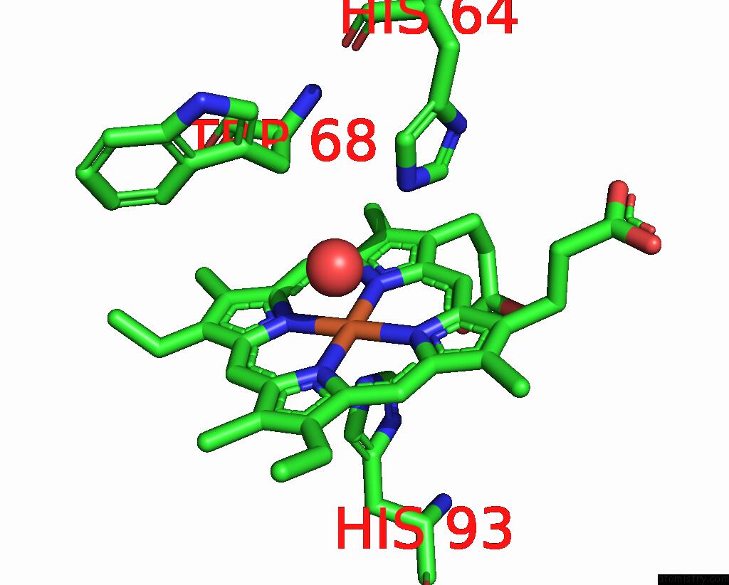
Mono view
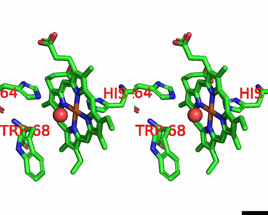
Stereo pair view
Mono view
Stereo pair view
A full contact list of Iron with other atoms in the Fe binding
site number 1 of Myoglobin Cavity Mutant V68W within 5.0Å range:
|
Reference:
J.S.Olson,
J.Soman,
G.N.Phillips.
Ligand Pathways in Myoglobin: A Review of Trp Cavity Mutations. Iubmb Life V. 59 552 2007.
ISSN: ISSN 1521-6543
PubMed: 17701550
DOI: 10.1080/15216540701230495
Page generated: Sun Aug 4 00:58:15 2024
ISSN: ISSN 1521-6543
PubMed: 17701550
DOI: 10.1080/15216540701230495
Last articles
Zn in 9MJ5Zn in 9HNW
Zn in 9G0L
Zn in 9FNE
Zn in 9DZN
Zn in 9E0I
Zn in 9D32
Zn in 9DAK
Zn in 8ZXC
Zn in 8ZUF