Iron »
PDB 2orl-2pg7 »
2owj »
Iron in PDB 2owj: Structure of An Early-Microsecond Photolyzed State of Co- Bjfixlh, Dark State
Protein crystallography data
The structure of Structure of An Early-Microsecond Photolyzed State of Co- Bjfixlh, Dark State, PDB code: 2owj
was solved by
J.Key,
V.Srajer,
R.Pahl,
K.Moffat,
with X-Ray Crystallography technique. A brief refinement statistics is given in the table below:
| Resolution Low / High (Å) | 34.10 / 2.50 |
| Space group | H 3 2 |
| Cell size a, b, c (Å), α, β, γ (°) | 128.500, 128.500, 58.400, 90.00, 90.00, 120.00 |
| R / Rfree (%) | 21.3 / 29.1 |
Iron Binding Sites:
The binding sites of Iron atom in the Structure of An Early-Microsecond Photolyzed State of Co- Bjfixlh, Dark State
(pdb code 2owj). This binding sites where shown within
5.0 Angstroms radius around Iron atom.
In total only one binding site of Iron was determined in the Structure of An Early-Microsecond Photolyzed State of Co- Bjfixlh, Dark State, PDB code: 2owj:
In total only one binding site of Iron was determined in the Structure of An Early-Microsecond Photolyzed State of Co- Bjfixlh, Dark State, PDB code: 2owj:
Iron binding site 1 out of 1 in 2owj
Go back to
Iron binding site 1 out
of 1 in the Structure of An Early-Microsecond Photolyzed State of Co- Bjfixlh, Dark State
Mono view
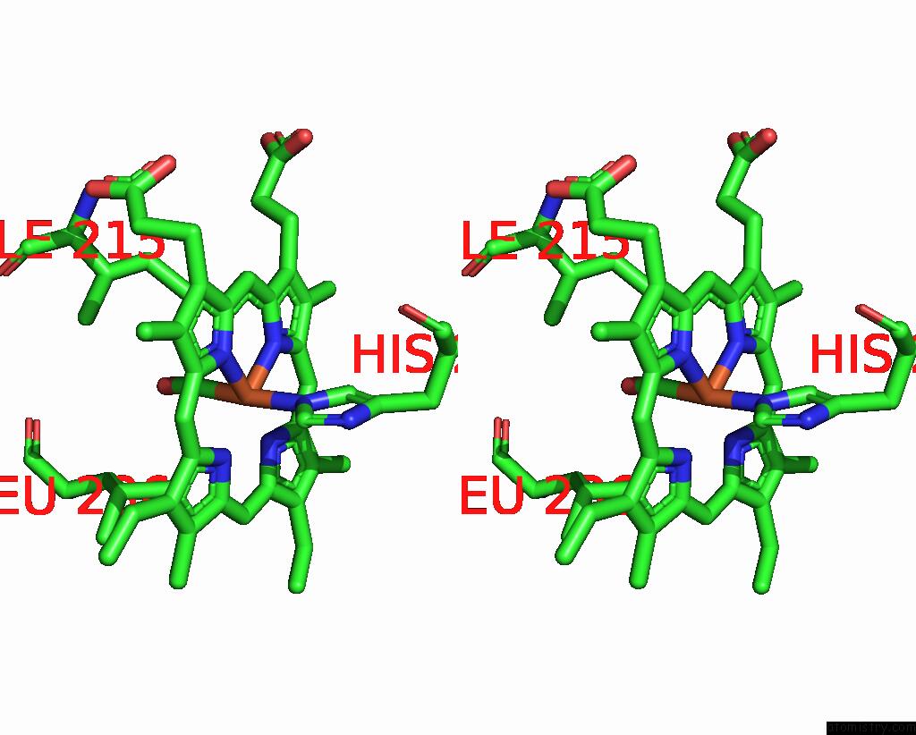
Stereo pair view
Mono view
Stereo pair view
A full contact list of Iron with other atoms in the Fe binding
site number 1 of Structure of An Early-Microsecond Photolyzed State of Co- Bjfixlh, Dark State within 5.0Å range:
|
Reference:
J.Key,
V.Srajer,
R.Pahl,
K.Moffat.
Time-Resolved Crystallographic Studies of the Heme Domain of the Oxygen Sensor Fixl: Structural Dynamics of Ligand Rebinding and Their Relation to Signal Transduction. Biochemistry V. 46 4706 2007.
ISSN: ISSN 0006-2960
PubMed: 17385895
DOI: 10.1021/BI700043C
Page generated: Thu Jul 17 03:16:52 2025
ISSN: ISSN 0006-2960
PubMed: 17385895
DOI: 10.1021/BI700043C
Last articles
Fe in 2YXOFe in 2YRS
Fe in 2YXC
Fe in 2YNM
Fe in 2YVJ
Fe in 2YP1
Fe in 2YU2
Fe in 2YU1
Fe in 2YQB
Fe in 2YOO