Iron »
PDB 2orl-2pg7 »
2pa8 »
Iron in PDB 2pa8: X-Ray Crystal Structure of the D/L Subcomplex of the Sulfolobus Solfataricus Rna Polymerase
Enzymatic activity of X-Ray Crystal Structure of the D/L Subcomplex of the Sulfolobus Solfataricus Rna Polymerase
All present enzymatic activity of X-Ray Crystal Structure of the D/L Subcomplex of the Sulfolobus Solfataricus Rna Polymerase:
2.7.7.6;
2.7.7.6;
Protein crystallography data
The structure of X-Ray Crystal Structure of the D/L Subcomplex of the Sulfolobus Solfataricus Rna Polymerase, PDB code: 2pa8
was solved by
A.Hirata,
K.S.Murakami,
with X-Ray Crystallography technique. A brief refinement statistics is given in the table below:
| Resolution Low / High (Å) | 15.00 / 1.76 |
| Space group | I 21 21 21 |
| Cell size a, b, c (Å), α, β, γ (°) | 69.703, 93.346, 128.277, 90.00, 90.00, 90.00 |
| R / Rfree (%) | 21 / 24.7 |
Iron Binding Sites:
The binding sites of Iron atom in the X-Ray Crystal Structure of the D/L Subcomplex of the Sulfolobus Solfataricus Rna Polymerase
(pdb code 2pa8). This binding sites where shown within
5.0 Angstroms radius around Iron atom.
In total 3 binding sites of Iron where determined in the X-Ray Crystal Structure of the D/L Subcomplex of the Sulfolobus Solfataricus Rna Polymerase, PDB code: 2pa8:
Jump to Iron binding site number: 1; 2; 3;
In total 3 binding sites of Iron where determined in the X-Ray Crystal Structure of the D/L Subcomplex of the Sulfolobus Solfataricus Rna Polymerase, PDB code: 2pa8:
Jump to Iron binding site number: 1; 2; 3;
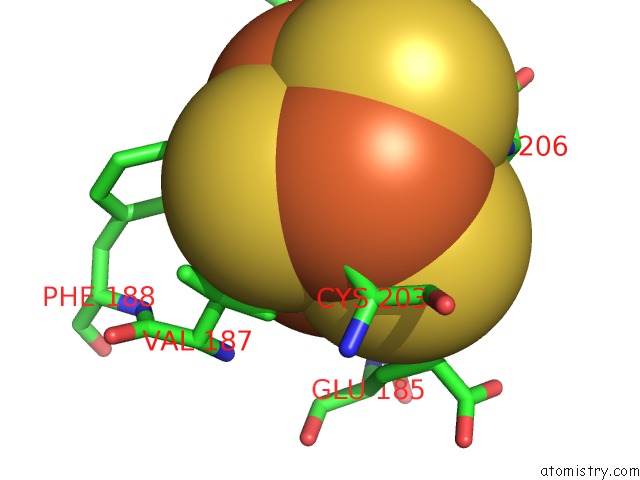
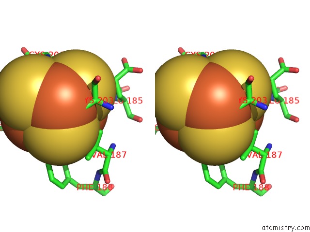
Iron binding site 1 out of 3 in 2pa8
Go back to
Iron binding site 1 out
of 3 in the X-Ray Crystal Structure of the D/L Subcomplex of the Sulfolobus Solfataricus Rna Polymerase
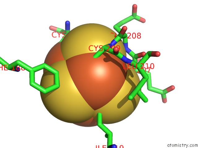
Mono view
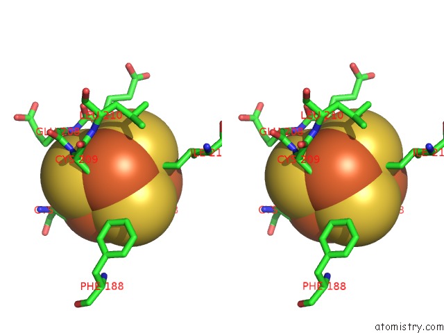
Stereo pair view
Mono view
Stereo pair view
A full contact list of Iron with other atoms in the Fe binding
site number 1 of X-Ray Crystal Structure of the D/L Subcomplex of the Sulfolobus Solfataricus Rna Polymerase within 5.0Å range:
|
Iron binding site 2 out of 3 in 2pa8
Go back to
Iron binding site 2 out
of 3 in the X-Ray Crystal Structure of the D/L Subcomplex of the Sulfolobus Solfataricus Rna Polymerase
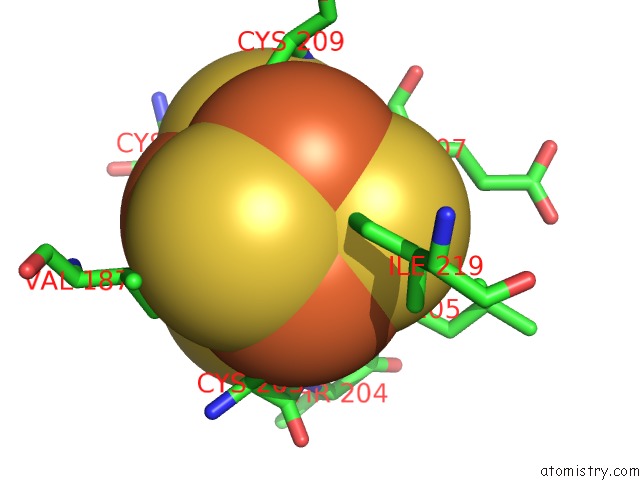
Mono view
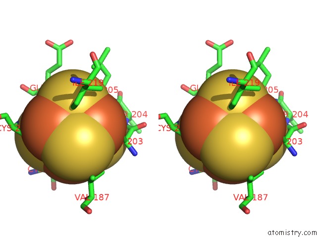
Stereo pair view
Mono view
Stereo pair view
A full contact list of Iron with other atoms in the Fe binding
site number 2 of X-Ray Crystal Structure of the D/L Subcomplex of the Sulfolobus Solfataricus Rna Polymerase within 5.0Å range:
|
Iron binding site 3 out of 3 in 2pa8
Go back to
Iron binding site 3 out
of 3 in the X-Ray Crystal Structure of the D/L Subcomplex of the Sulfolobus Solfataricus Rna Polymerase
Mono view
Stereo pair view
Mono view
Stereo pair view
A full contact list of Iron with other atoms in the Fe binding
site number 3 of X-Ray Crystal Structure of the D/L Subcomplex of the Sulfolobus Solfataricus Rna Polymerase within 5.0Å range:
|
Reference:
A.Hirata,
B.J.Klein,
K.S.Murakami.
The X-Ray Crystal Structure of Rna Polymerase From Archaea. Nature V. 451 851 2008.
ISSN: ISSN 0028-0836
PubMed: 18235446
DOI: 10.1038/NATURE06530
Page generated: Thu Jul 17 03:22:20 2025
ISSN: ISSN 0028-0836
PubMed: 18235446
DOI: 10.1038/NATURE06530
Last articles
Fe in 2YXOFe in 2YRS
Fe in 2YXC
Fe in 2YNM
Fe in 2YVJ
Fe in 2YP1
Fe in 2YU2
Fe in 2YU1
Fe in 2YQB
Fe in 2YOO