Iron »
PDB 2orl-2pg7 »
2pbj »
Iron in PDB 2pbj: Gsh-Heme Bound Microsomal Prostaglandin E Synthase
Enzymatic activity of Gsh-Heme Bound Microsomal Prostaglandin E Synthase
All present enzymatic activity of Gsh-Heme Bound Microsomal Prostaglandin E Synthase:
5.3.99.3;
5.3.99.3;
Protein crystallography data
The structure of Gsh-Heme Bound Microsomal Prostaglandin E Synthase, PDB code: 2pbj
was solved by
F.Takusagawa,
T.Yamada,
with X-Ray Crystallography technique. A brief refinement statistics is given in the table below:
| Resolution Low / High (Å) | 20.00 / 2.80 |
| Space group | C 1 2 1 |
| Cell size a, b, c (Å), α, β, γ (°) | 127.000, 122.490, 110.970, 90.00, 111.00, 90.00 |
| R / Rfree (%) | 24.2 / 26.9 |
Other elements in 2pbj:
The structure of Gsh-Heme Bound Microsomal Prostaglandin E Synthase also contains other interesting chemical elements:
| Chlorine | (Cl) | 4 atoms |
Iron Binding Sites:
The binding sites of Iron atom in the Gsh-Heme Bound Microsomal Prostaglandin E Synthase
(pdb code 2pbj). This binding sites where shown within
5.0 Angstroms radius around Iron atom.
In total 4 binding sites of Iron where determined in the Gsh-Heme Bound Microsomal Prostaglandin E Synthase, PDB code: 2pbj:
Jump to Iron binding site number: 1; 2; 3; 4;
In total 4 binding sites of Iron where determined in the Gsh-Heme Bound Microsomal Prostaglandin E Synthase, PDB code: 2pbj:
Jump to Iron binding site number: 1; 2; 3; 4;
Iron binding site 1 out of 4 in 2pbj
Go back to
Iron binding site 1 out
of 4 in the Gsh-Heme Bound Microsomal Prostaglandin E Synthase
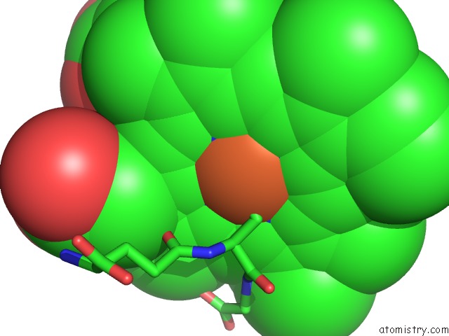
Mono view

Stereo pair view
Mono view
Stereo pair view
A full contact list of Iron with other atoms in the Fe binding
site number 1 of Gsh-Heme Bound Microsomal Prostaglandin E Synthase within 5.0Å range:
|
Iron binding site 2 out of 4 in 2pbj
Go back to
Iron binding site 2 out
of 4 in the Gsh-Heme Bound Microsomal Prostaglandin E Synthase
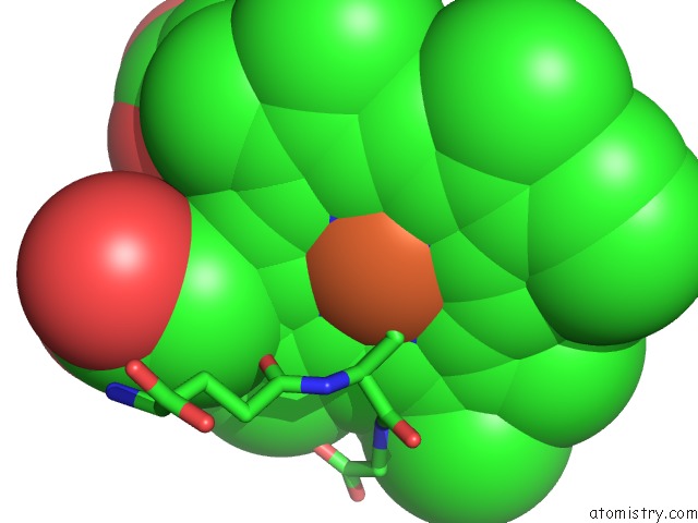
Mono view
Stereo pair view
Mono view
Stereo pair view
A full contact list of Iron with other atoms in the Fe binding
site number 2 of Gsh-Heme Bound Microsomal Prostaglandin E Synthase within 5.0Å range:
|
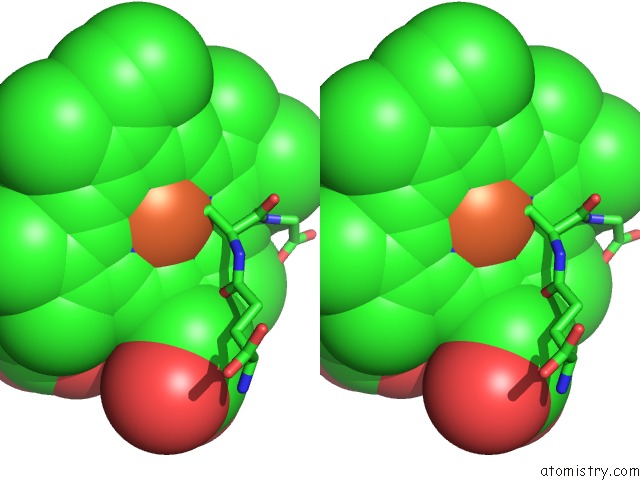
Iron binding site 3 out of 4 in 2pbj
Go back to
Iron binding site 3 out
of 4 in the Gsh-Heme Bound Microsomal Prostaglandin E Synthase
Mono view

Stereo pair view
Mono view
Stereo pair view
A full contact list of Iron with other atoms in the Fe binding
site number 3 of Gsh-Heme Bound Microsomal Prostaglandin E Synthase within 5.0Å range:
|
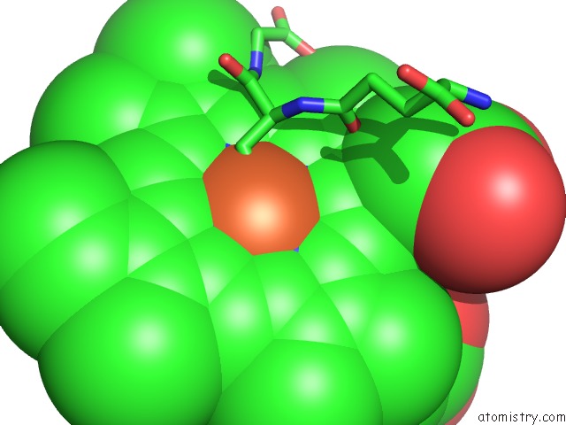
Iron binding site 4 out of 4 in 2pbj
Go back to
Iron binding site 4 out
of 4 in the Gsh-Heme Bound Microsomal Prostaglandin E Synthase
Mono view

Stereo pair view
Mono view
Stereo pair view
A full contact list of Iron with other atoms in the Fe binding
site number 4 of Gsh-Heme Bound Microsomal Prostaglandin E Synthase within 5.0Å range:
|
Reference:
T.Yamada,
F.Takusagawa.
PGH2 Degradation Pathway Catalyzed By Gsh-Heme Complex Bound Microsomal Prostaglandin E2 Synthase Type 2: the First Example of A Dual-Function Enzyme. Biochemistry V. 46 8414 2007.
ISSN: ISSN 0006-2960
PubMed: 17585783
DOI: 10.1021/BI700605M
Page generated: Thu Jul 17 03:23:28 2025
ISSN: ISSN 0006-2960
PubMed: 17585783
DOI: 10.1021/BI700605M
Last articles
Fe in 2YXOFe in 2YRS
Fe in 2YXC
Fe in 2YNM
Fe in 2YVJ
Fe in 2YP1
Fe in 2YU2
Fe in 2YU1
Fe in 2YQB
Fe in 2YOO